

ω-3 Tear Film Lipids Correlate With Clinical Measures of Dry Eye
- PMID: 27138739
- PMCID: PMC4857833
- DOI: 10.1167/iovs.16-19131
ω-3 Tear Film Lipids Correlate With Clinical Measures of Dry Eye
Abstract
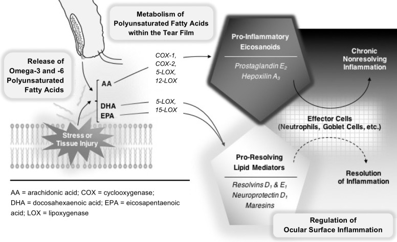
Purpose: ω-3 and ω-6 polyunsaturated fatty acids modulate inflammatory processes throughout the body through distinct classes of lipid mediators that possess both proinflammatory and proresolving properties. The purpose of this cross-sectional study was to explore the relationship between lipid profiles in human tears and dry eye (DE) symptoms and signs.
Methods: Forty-one patients with normal eyelid and corneal anatomy were prospectively recruited from a Veterans Administration Hospital over 18 months. Symptoms and signs of DE were assessed, and tear samples was analyzed by mass spectrometry-based lipidomics. Statistical analyses comparing the relationship between tear film lipids and DE included Pearson/Spearman correlations and t-tests.
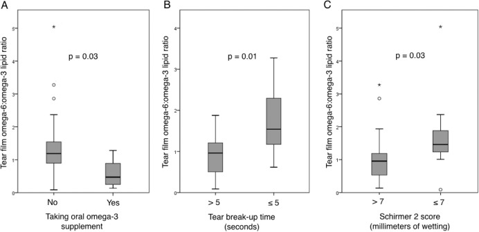
Results: Arachidonic acid (AA), docosahexaenoic acid (DHA), and eicosapentaenoic acid (EPA) were present in more than 90% of tear film samples. The ratio of ω-6 (AA) to ω-3 (DHA+EPA) fatty acids was correlated with multiple measures of tear film dysfunction (tear breakup time, Schirmer 2 scores, and corneal staining; all P < 0.05). Arachidonic acid-derived prostaglandin E2 was detected in the majority of samples and correlated with low tear osmolarity, meibomian gland plugging, and corneal staining.
Conclusions: Both ω-3 and ω-6 lipid circuits are activated in the human tear film. The ratio of ω-6:ω-3 tear lipids is elevated in DE patients in proportion to the degree of tear film dysfunction and corneal staining. Metabolic deficiency of ω-3 tear film lipids may be a driver of chronic ocular surface inflammation in DE.
Figures
References
-
- The epidemiology of dry eye disease: report of the Epidemiology Subcommittee of the International Dry Eye WorkShop (2007). Ocul Surf. 2007; 5: 93–107. - PubMed
-
- Pouyeh B,, Viteri E,, Feuer W,, et al. Impact of ocular surface symptoms on quality of life in a United States Veterans Affairs population. Am J Ophthalmol. 2012; 153: 1061–1066. - PubMed
-
- Stern ME,, Gao J,, Schwalb TA,, et al. Conjunctival T-cell subpopulations in Sjogren's and non-Sjogren's patients with dry eye. Invest Ophthalmol Vis Sci. 2002; 43: 2609–2614. - PubMed
Publication types
MeSH terms
Substances
Grants and funding
LinkOut - more resources
Full Text Sources
Other Literature Sources
Medical
Research Materials
