. 2016 May 3:7:11430.
doi: 10.1038/ncomms11430.

Wnt pathway activation by ADP-ribosylation

Affiliations

Wnt pathway activation by ADP-ribosylation

Eungi Yang et al. Nat Commun. .

Abstract

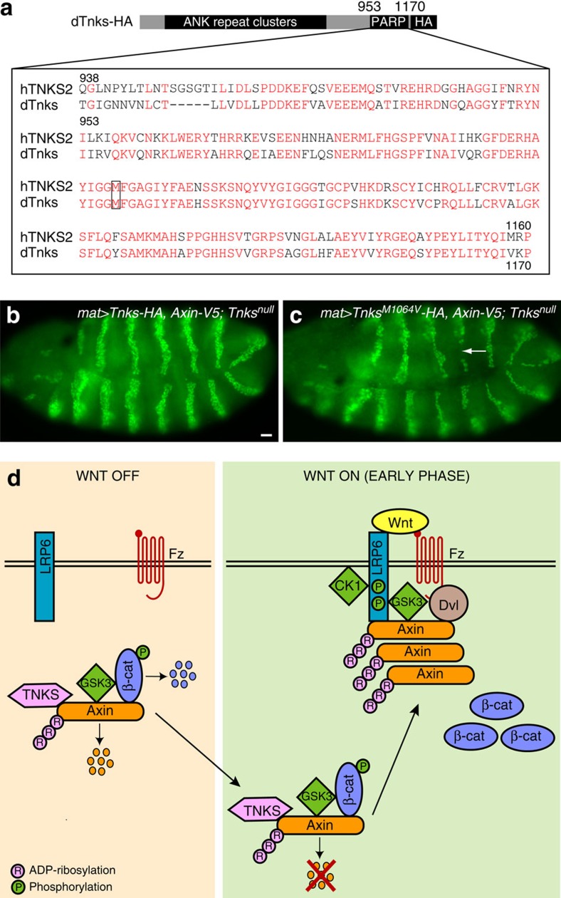
Wnt/β-catenin signalling directs fundamental processes during metazoan development and can be aberrantly activated in cancer. Wnt stimulation induces the recruitment of the scaffold protein Axin from an inhibitory destruction complex to a stimulatory signalosome. Here we analyse the early effects of Wnt on Axin and find that the ADP-ribose polymerase Tankyrase (Tnks)--known to target Axin for proteolysis-regulates Axin's rapid transition following Wnt stimulation. We demonstrate that the pool of ADP-ribosylated Axin, which is degraded under basal conditions, increases immediately following Wnt stimulation in both Drosophila and human cells. ADP-ribosylation of Axin enhances its interaction with the Wnt co-receptor LRP6, an essential step in signalosome assembly. We suggest that in addition to controlling Axin levels, Tnks-dependent ADP-ribosylation promotes the reprogramming of Axin following Wnt stimulation; and propose that Tnks inhibition blocks Wnt signalling not only by increasing destruction complex activity, but also by impeding signalosome assembly.


Figures

Figure 1
Figure 1. Rapid appearance of Axin in segmental stripes following Wg exposure.
(al) Axin is distributed uniformly in the embryonic ectoderm before and at the onset of Wingless (Wg) expression. Confocal images of embryos expressing Axin-V5 driven by the mat-Gal4 driver. Genotype left, antibodies top. Embryonic stage and developmental time in hours after egg lay (AEL) are indicated at the top right of a,g and m. Anterior left, dorsal up. In stage 5 and 6 embryos, Axin-V5 and the transmembrane protein Neurotactin are distributed uniformly throughout the ectoderm (al). Higher magnification images (df and jl). (mr) Axin is distributed in segmental stripes after the onset of Wg expression. By stage 9, Axin-V5 accumulates in segmental stripes with increased staining intensity (mo). Higher magnification images (pr). White bars indicate the width of a single Axin stripe. Scale bar, 25 μm.
Figure 2
Figure 2. Axin is rapidly stabilized and subsequently destabilized following Wg exposure.
(ac) Axin is distributed uniformly in the ectoderm at the onset of Wg exposure. Embryos expressing Axin-V5 driven by mat-Gal4 (mat>Axin-V5) stained with V5 and Wg antibodies. Stage of embryonic development and corresponding time relative to the onset of Wg expression is indicated at bottom left of c,f,i,l. At 3 h of development (stage 6), Axin-V5 is distributed uniformly throughout the embryonic ectoderm and the initial expression of Wg in segmental stripes is weak. (df) Axin levels increase rapidly following Wg exposure. By 30 to 40 min after the onset of Wg expression (late stage 8 to stage 9), Axin-V5 is distributed in wide segmental stripes (indicated by brackets) that overlap the narrow Wg stripes (asterisks). (gl) Axin levels subsequently decrease, beginning at 2 h after Wg exposure. (gi) Approximately 120 min after onset of Wg expression, mid-stage 10 embryos. During this transitional stage, the Axin-V5 level is decreased precisely at the position of the Wg stripes (asterisks), but remains elevated in the neighbouring cells (brackets). (jl) Embryos expressing Axin-V5 driven by arm-Gal4 (arm>Axin-V5) stained with V5 and Wg antibodies. arm-Gal4 drives ubiquitous transcription at late embryonic stages, and thus allows analysis of Axin regulation in stage 11 embryos. By 240 min after the onset of Wg exposure, the Axin-V5 level is markedly decreased at the position of the Wg stripes. Wg (asterisks) and Axin stripes are spatially juxtaposed. (c*–l*) Schematic illustration of the spatial relationship between Axin-V5 and Wg with respect to the onset of Wg exposure. (mr) Axin levels increase rapidly in response to Wg exposure. Confocal images of stage 9 embryo (mo) and higher magnification images of boxed region (pr). Axin stripes (white bars) are centered directly over Wg expressing cells (arrows). (sx) Axin levels are decreased by several hours after Wg exposure. Confocal images of stage 13 embryo expressing Axin-V5 with the arm-Gal4 driver. By this time, Axin and Wg stripes are spatially juxtaposed (su), as shown in higher magnification images (vx). Asterisks indicate the position of Wg stripe. For all images, anterior left, dorsal up. Scale bar, 25 μm.
Figure 3
Figure 3. Tnks promotes Axin regulation during the early phase, but is dispensable for Axin proteolysis during the delayed phase after Wg exposure.
(a-f) Wild-type Axin is initially stabilized and subsequently degraded following Wg exposure. Stage 9 and mid-stage 10 embryos expressing Axin-V5 driven by the mat-Gal4 driver, co-stained with V5 and Wg antibodies. Genotypes left, antibodies top. (ac) By 40 min after the onset of Wg exposure (stage 9), Axin-V5 is distributed in wide segmental stripes (brackets) that overlap narrow Wg stripes (asterisks). (df) By 120 min after the onset of Wg exposure (mid-stage 10), Axin-V5 staining is decreased in cells expressing Wg (asterisks). (gl) The Tnks-binding domain in Axin is required for the initial stabilization of Axin induced by Wg exposure, but dispensable for the subsequent Wg-dependent Axin proteolysis. (gi) In stage 9 embryos, AxinΔTBD-V5 staining is uniformly high throughout the embryonic ectoderm; no segmental stripes are present. (jl) In mid-stage 10 embryos, the AxinΔTBD-V5 levels are decreased in cells expressing Wg (asterisks). Axin stripes are spatially juxtaposed with Wg stripes. (mr) Tnks is required for the initial stabilization of Axin induced by Wg exposure, but dispensable for the subsequent Wg-dependent Axin proteolysis. (mo) In stage 9 Tnks null mutant embryos, Axin-V5 staining is uniformly high in all ectodermal cells. (pr) In mid-stage 10 Tnks null mutant embryos, Axin-V5 is decreased in cells expressing Wg (asterisks). Axin stripes are spatially juxtaposed with Wg stripes. Scale bar, 25 μm. Schematic illustration of spatial relationship of Axin-V5 and Wg with respect to the onset of Wg expression in c*–r*.
Figure 4
Figure 4. Tnks promotes the activation of Wg signalling.
Tnks and the TBD in Axin promote Wg signalling. Confocal images of stage 8 or 9 embryos stained with Engrailed (En) antibody. Genotypes left, developmental stage indicated on top. (a,b) In wild-type embryos expressing Axin-V5, En is present in stripes that are 2 to 3 cells in width (b*, white bar), at both 10 min after the onset of Wg expression (stage 8) and 40 min after the onset of Wg (stage 9). (c,d) In embryos expressing AxinΔTBD-V5, the initiation of En stripes (stage 8) is normal (c), but En stripes decay in 68% (n=50) of embryos (d), as revealed by the aberrantly narrowed width by stage 9 (d*, arrow). (e,f) In Tnks null mutant embryos expressing Axin-V5, En expression is normal at stage 8 (e), but decays by stage 9 in 75% (n=36) of embryos (f*, arrow). Images in b*,d* and f* are higher magnification views of embryos in b,d, and f respectively. In all images, anterior left, dorsal up. Scale bar, 25 μm.
Figure 5
Figure 5. In addition to its known role in Axin proteolysis, Tnks promotes Wg pathway activation through a novel mechanism of Axin regulation.
(a-b) Generation of transgenic flies with wild-type Axin at levels higher than those resulting from loss of Tnks-dependent Axin proteolysis. (a) Immunoblot analysis of lysates from embryos expressing either Axin-V5 or AxinΔTBD-V5 integrated at the attP33 or the attP40 site. Lysates were prepared from embryos collected at 0–2 h of development, before the onset of Wg expression. Levels of V5 from attP33 AxinΔTBD-V5 are higher than attP33 Axin-V5 when expressed under the same conditions. Integration of the wild-type Axin-V5 transgene at a different genomic site, attP40, results in levels that are higher than attP33 AxinΔTBD-V5. Kinesin was used as a loading control. (b) Quantification of relative levels of indicated protein from experiments shown in a. Error bars represent s.e.m. of three independent experiments. P=0.0348 (one-way ANOVA with Kruskal–Wallis test). (c) The hatch rate of embryos expressing attP40 Axin-V5, attP33 Axin-V5 or attP33 AxinΔTBD-V5 with the mat-Gal4 driver. By comparison with embryos expressing attP33 Axin-V5 or attP40 Axin-V5, the hatch rate is reduced by expression of attp33 AxinΔTBD-V5. At least 200 embryos of each genotype were analysed, as indicated. (di) An increase in wild-type Axin, above the levels resulting from loss of Tnks-dependent Axin proteolysis, is compatible with the rapid stabilization of Axin following Wg exposure. (df) In stage 9 embryos expressing attP40 Axin-V5 driven by mat-Gal4, Axin-V5 accumulates in segmental stripes (brackets) that overlap the Wg stripes (asterisks). (gi) Co-staining with V5 and Arm antibodies shows that Axin-V5 accumulates normally in Wg-responding cells, as revealed by co-localization with cells that accumulate Arm (white bars). (j,k) An increase in wild-type Axin, above the levels resulting from loss of Tnks-dependent Axin proteolysis, is compatible with the activation of Wg signalling. En expression is normal at both stage 8 (j) and stage 9 (k). A higher magnification view of the En stripes in k is shown (k*, white bar indicates the width of a single En stripe). Scale bar, 25 μm.
Figure 6
Figure 6. ADP-ribosylated Axin rapidly accumulates following Wnt stimulation.
(a) Use of a GST-WWE pull down assay for specific detection of ADP-ribosylated Axin. Detection of ADP-ribosylated mouse Flag-Axin1 and Flag-Axin1ΔTBD using either GST-WWE or GST-WWER163A control pull downs in HEK293T cells. Flag-Axin1 is pulled down by GST-WWE; however, the Flag-Axin1ΔTBD control is not. (b,c) The levels of ADP-ribosylated Axin rapidly increased following Wnt exposure in human cells. (b) HEK293T cells were treated with Wnt3A for 0.5 h, and cell lysates were subsequently pulled down by GST-WWE followed by immunoblot analysis with the indicated antibodies. Following treatment with Wnt, phospho-LRP6 is pulled down by GST-WWE. (c) Quantification of relative ADP-ribosylated Axin1 levels from experiments shown in (b). Error bars represent s.e.m. of three independent experiments. P=0.0062 (Student's t-test). (d,e) The level of ADP-ribosylated Axin rapidly increased following Wg exposure in Drosophila cells. (d) Immunoblot of lysates from S2R+ cells treated with Wg conditioned medium (CM) for 1 h and then subjected to GST-WWE pull down. Treatment with Wg CM increased the level of ADP-ribosylated Axin pulled down with GST-WWE. Following treatment with Wg CM, phospho-Arrow is pulled down by GST-WWE. (d, input) Treatment with Wg CM induced a mobility shift in Axin. (e) Quantification of relative levels of ADP-ribosylated Axin from experiments shown in (d). Error bars represent s.e.m. of four independent experiments. P=0.0147 (Student's t-test). (fh) The levels of Axin-V5 and ADP-ribosylated Axin-V5 are rapidly increased after the onset of Wg stimulation in Drosophila embryos. Embryos at stages 4–5 and stage 9 expressing Axin-V5 driven by the mat-Gal4 driver. Stages 4-5 (1.5 to 2.5 h of development) are before the onset of Wg stimulation and stage 9 (3.5 to 4.5 h of development) is after the onset of Wg stimulation. Lysates were immunoblotted by indicated antibodies, followed by GST-WWE pull down. Axin-V5 and phospho-Arrow are pulled down by GST-WWE following Wg stimulation. (g and h) Quantification of relative Axin-V5 and ADP-ribosylated Axin-V5 levels from experiments shown in (f). (g) P=0.0321 (Student's t-test). (h) P=0.0305 (Student's t-test). Error bars represent s.e.m. of three independent experiments.
Figure 7
Figure 7. ADP-ribosylation enhances interaction of Axin with phospho-LRP6 following Wnt stimulation.
(a) Phospho-Arrow interacts with ADP-ribosylated Axin following Wg exposure. Immunoblot of S2R+ cells treated with dsRNA against Axin or white (negative control), then treated with Wg CM for 1 h, subjected to GST-WWE pull down, and analysed by immunoblotting with indicated antibodies. Treatment with Wg CM markedly increases the amount of ADP-ribosylated Axin pulled down with GST-WWE, but not the GST-WWER163A control. On treatment with Wg CM, phospho-Arrow is also pulled down with GST-WWE, but not the GST-WWER163A control. The pull down of phosphorylated Arrow by GST-WWE is prevented by treatment with Axin dsRNA. (a, input) Treatment with Wg CM induced a mobility shift in Axin. There is a small decrease in the level of phospho-LRP6 following Axin knockdown. These data suggest that in Drosophila, Axin is not essential for Wg-dependent phosphorylation of LRP6, but may facilitate this process. (b) ADP-ribosylation enhances the interaction of Axin with phospho-LRP6. HEK293T cells expressing Flag-Axin1 were treated with either the DMSO control or the small molecule Tnks inhibitor XAV939 for 24 h. XAV939 diminishes the Wnt3A-dependent interaction between Axin1 and phospho-LRP6. (c,d) The Tnks-binding domain enhances the interaction of Axin with phospho-LRP6 following Wnt exposure. HEK293T cells transfected with either Flag-Axin1 or Flag-Axin1ΔTBD, and subsequently treated with Wnt3A for 30 min. Lysates were immunoprecipitated by Flag antibody, followed by immunoblot using indicated antibodies. Both (c) phospho-LRP6 (S1490) and (d) phospho-LRP6 (T1572) antibodies demonstrate that Axin1 interacts with phospho-LRP6 following Wnt3A stimulation; however, deletion of the TBD of Axin1 diminishes this interaction. α-Tub was used as a loading control. These experiments were performed at least three times with representative results shown.
Figure 8
Figure 8. Tnks catalytic activity promotes activation of the Wg pathway.
(a) Schematic representation of HA-tagged Drosophila Tnks (dTnks-HA). Alignment below shows amino-acid conservation in the catalytic PARP domain of human and Drosophila Tnks. Identical amino acids are in red. Box indicates the substitution in dTnksM1064V that is predicted to inactivate PARP activity. (b,c) Wg signalling in the Tnks null mutant is rescued by expression of wild-type Tnks, but not catalytically inactive Tnks. Stage 9 embryos stained with En antibody. (b) In Tnks null mutant embryos expressing Tnks-HA and Axin-V5 driven by the mat-Gal4 driver, the width of the En stripes is normal (2–3 cells in width). (c) In Tnks null mutant embryos expressing TnksM1064V-HA and Axin-V5 driven by the mat-Gal4 driver, En stripes are aberrantly narrowed (arrow). Scale bar, 25 μm. (d) Model for the dual roles of Tnks in Wnt signalling. Tnks-dependent ADP-ribosylation not only targets Axin for proteolysis independently of Wnt stimulation, but also promotes signalling immediately following Wnt exposure. Wnt stimulation induces the rapid accumulation of ADP-ribosylated Axin. ADP-ribosylation promotes Axin's interaction with the Wnt co-receptor LRP6, thereby activating the Wnt pathway.

References

    1. MacDonald B. T., Tamai K. & He X. Wnt/beta-catenin signaling: components, mechanisms, and diseases. Dev. Cell. 17, 9–26 (2009). - PMC - PubMed
    1. Clevers H. & Nusse R. Wnt/beta-catenin signaling and disease. Cell 149, 1192–1205 (2012). - PubMed
    1. Ikeda S. et al.. Axin, a negative regulator of the Wnt signaling pathway, forms a complex with GSK-3beta and beta-catenin and promotes GSK-3beta-dependent phosphorylation of beta-catenin. EMBO J. 17, 1371–1384 (1998). - PMC - PubMed
    1. Behrens J. et al.. Functional interaction of an axin homolog, conductin, with beta-catenin, APC, and GSK3beta. Science 280, 596–599 (1998). - PubMed
    1. Kimelman D. & Xu W. beta-catenin destruction complex: insights and questions from a structural perspective. Oncogene 25, 7482–7491 (2006). - PubMed

Publication types

MeSH terms

LinkOut - more resources