. 2016 Aug;140(8):836-43.
doi: 10.5858/arpa.2015-0220-OA. Epub 2016 May 3.

Interobserver Variability in the Diagnosis of Uterine High-Grade Endometrioid Carcinoma

Affiliations

Interobserver Variability in the Diagnosis of Uterine High-Grade Endometrioid Carcinoma

Sumi Thomas et al. Arch Pathol Lab Med. 2016 Aug.

Abstract

Context: -Low interobserver diagnostic agreement exists among high-grade endometrial carcinomas.

Objective: -To evaluate diagnostic variability in International Federation of Gynecology and Obstetrics (FIGO) grade 3 endometrioid adenocarcinoma (G3EC) in 2 different sign-out practices.

Design: -Sixty-six G3EC cases were identified from pathology archives of Wayne State University (WSU, Detroit, Michigan) (general surgical pathology sign-out) and 65 from Memorial Sloan Kettering Cancer Center (MSK, New York, New York) (gynecologic pathology focused sign-out). Each case was reviewed together by 2 gynecologic pathologists, one from each institution, and classified into the G3EC group or a reclassified group. Clinicopathologic parameters were compared.

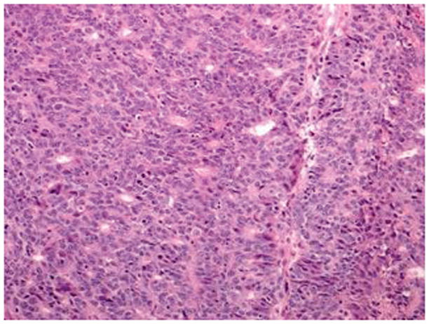
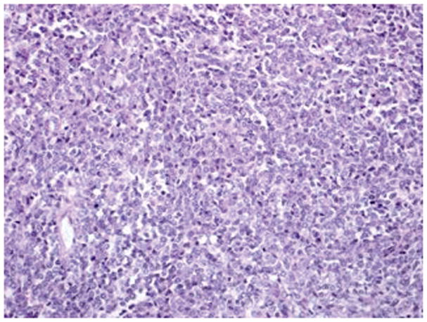
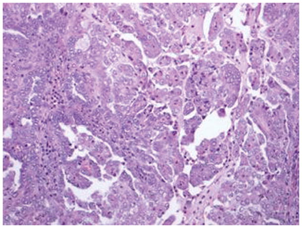
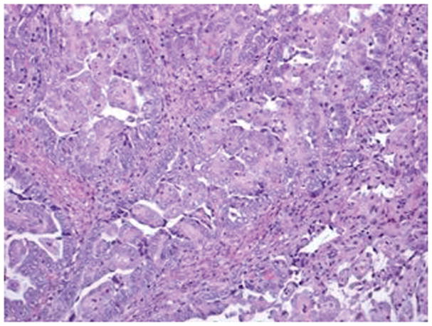
Results: -Twenty-five WSU cases (38%) were reclassified as undifferentiated (n = 2), serous (n = 4), mixed endometrioid and serous carcinomas (n = 12), and FIGO grade 2 endometrioid adenocarcinomas with focal marked nuclear atypia (n = 7). Eleven MSK cases (17%) were reclassified as undifferentiated (n = 5), serous (n = 1), mixed endometrioid and serous carcinomas (n = 4), and mixed endometrioid and clear cell carcinomas (n = 1). Agreement rate between original and review diagnosis was 83% (54 of 65) at MSK and 62% (41 of 66) at WSU (P = .01) with an overall rate of 73% (95 of 131). There were more undifferentiated carcinomas at MSK than there were at WSU (45% [5 of 11] versus 8% [2 of 25]; P = .02). There were more grade 2 endometrioid adenocarcinomas with focal, marked nuclear atypia at WSU (28%; 7 of 25) than there were at MSK (0%) (P = .03). Mixed endometrioid and serous carcinoma was the most common misclassified subtype (44%; 16 of 36).

Conclusion: -Moderate interobserver variability exists in the diagnosis of G3EC with a significantly greater diagnostic agreement rate in gynecologic pathology-focused sign-out than in general sign-out practice.


Figures

Figure 1
Figure 1
FIGO grade 3 endometrioid carcinoma has >50 % solid architecture with focal gland formation and moderate nuclear atypia (hematoxylin-eosin, original magnification ×20)
Figure 2
Figure 2
Undifferentiated carcinoma has monotonous proliferation of medium sized cells in solid and diffuse pattern (hematoxylin-eosin, original magnification ×20)
Figure 3
Figure 3
Serous carcinoma features a papillary component with slit like spaces (A, B) and/or a solid component (C, D) with high nuclear grade. Pleomorphic nuclei with prominent nucleoli, hyperchromatic nuclei with smudged chromatin and increased mitotic activity are readily observed (hematoxylin-eosin, original magnifications ×20 [A, B, C and D]
Figure 3
Figure 3
Serous carcinoma features a papillary component with slit like spaces (A, B) and/or a solid component (C, D) with high nuclear grade. Pleomorphic nuclei with prominent nucleoli, hyperchromatic nuclei with smudged chromatin and increased mitotic activity are readily observed (hematoxylin-eosin, original magnifications ×20 [A, B, C and D]
Figure 4
Figure 4
Mixed epithelial carcinoma: Areas of high-grade endometrioid carcinoma showing predominantly solid growth, focal evidence of gland formation and moderate nuclear atypia (A). Other areas in the same case have serous carcinoma (B) with papillary/glandular architecture, high nuclear grade, increased mitosis and necrotic luminal debris (hematoxylin-eosin, original magnifications ×20 [A, B]).
Figure 4
Figure 4
Mixed epithelial carcinoma: Areas of high-grade endometrioid carcinoma showing predominantly solid growth, focal evidence of gland formation and moderate nuclear atypia (A). Other areas in the same case have serous carcinoma (B) with papillary/glandular architecture, high nuclear grade, increased mitosis and necrotic luminal debris (hematoxylin-eosin, original magnifications ×20 [A, B]).
Figure 5
Figure 5
FIGO grade 2 endometrioid carcinoma with focal nuclear atypia: Area of endometrioid carcinoma showing low nuclear grade with oval nuclei and inconspicuous nucleoli (A) and foci in the same case showing higher nuclear grade with rounded nuclei, open chromatin, and distinct nucleoli (B) (hematoxylin-eosin, original magnifications ×20 [A, B]).
Figure 5
Figure 5
FIGO grade 2 endometrioid carcinoma with focal nuclear atypia: Area of endometrioid carcinoma showing low nuclear grade with oval nuclei and inconspicuous nucleoli (A) and foci in the same case showing higher nuclear grade with rounded nuclei, open chromatin, and distinct nucleoli (B) (hematoxylin-eosin, original magnifications ×20 [A, B]).

References

    1. Bokhman JV. Two pathogenetic types of endometrial carcinoma. Gynecol Oncol. 1983;15(1):10–17. - PubMed
    1. Hamilton CA, Cheung MK, Osann K, et al. Uterine papillary serous and clear cell carcinomas predict for poorer survival compared to grade 3 endometrioid corpus cancers. Br J Cancer. 2006;94(5):642–646. - PMC - PubMed
    1. Voss MA, Ganesan R, Ludeman L, et al. Should grade 3 endometrioid endometrial carcinoma be considered a type 2 cancer-a clinical and pathological evaluation. Gynecol Oncol. 2012;124(1):15–20. - PubMed
    1. Soslow RA, Bissonnette JP, Wilton A, et al. Clinicopathologic analysis of 187 high-grade endometrial carcinomas of different histologic subtypes: similar outcomes belie distinctive biologic differences. Am J Surg Pathol. 2007;31(7):979–987. - PubMed
    1. Creasman WT, Kohler MF, Odicino F, et al. Prognosis of papillary serous, clear cell, and grade 3 stage I carcinoma of the endometrium. Gynecol Oncol. 2004;95(3):593–596. - PubMed