. 2016 Jun 20;60(7):4206-16.
doi: 10.1128/AAC.03130-15. Print 2016 Jul.

Antifungal Mechanism of Action of Lactoferrin: Identification of H+-ATPase (P3A-Type) as a New Apoptotic-Cell Membrane Receptor

Affiliations

Antifungal Mechanism of Action of Lactoferrin: Identification of H+-ATPase (P3A-Type) as a New Apoptotic-Cell Membrane Receptor

María T Andrés et al. Antimicrob Agents Chemother. .

Abstract

Human lactoferrin (hLf) is a protein of the innate immune system which induces an apoptotic-like process in yeast. Determination of the susceptibility to lactoferrin of several yeast species under different metabolic conditions, respiratory activity, cytoplasmic ATP levels, and external medium acidification mediated by glucose assays suggested plasma membrane Pma1p (P3A-type ATPase) as the hLf molecular target. The inhibition of plasma membrane ATPase activity by hLf and the identification of Pma1p as the hLf-binding membrane protein confirmed the previous physiological evidence. Consistent with this, cytoplasmic ATP levels progressively increased in hLf-treated Candida albicans cells. However, oligomycin, a specific inhibitor of the mitochondrial F-type ATPase proton pump (mtATPase), abrogated the antifungal activity of hLf, indicating a crucial role for mtATPase in the apoptotic process. We suggest that lactoferrin targeted plasma membrane Pma1p H(+)-ATPase, perturbing the cytoplasmic ion homeostasis (i.e., cytoplasmic H(+) accumulation and subsequent K(+) efflux) and inducing a lethal mitochondrial dysfunction. This initial event involved a normal mitochondrial ATP synthase activity responsible for both the ATP increment and subsequent hypothetical mitochondrial proton flooding process. We conclude that human lactoferrin inhibited Pma1p H(+)-ATPase, inducing an apoptotic-like process in metabolically active yeast. Involvement of mitochondrial H(+)-ATPase (nonreverted) was essential for the progress of this programmed cell death in which the ionic homeostasis perturbation seems to precede classical nonionic apoptotic events.


Figures

FIG 1
FIG 1
Influence of the energetic metabolism on the fungicidal activity of lactoferrin. (A) Viability of yeast cells (105 cells/ml) in Tris buffer incubated for 90 min at 37°C with three different concentrations of hLf under anoxic (white columns) and oxygenic (black columns) conditions. Assays under anoxic conditions were performed with starved yeast cells. (B) Viability of yeast cells grown in the presence of 2% glucose (fermentation) (white columns) or 2% glycerol (respiration) (black columns) incubated with hLf for 90 min at 37°C. C. albicans cells were incubated with the metabolic inhibitor 2-DG before the hLf exposition (dashed columns). Aliquots were plated and colonies were counted after 24 h. The results are the means ± SD from duplicates of at least three independent experiments. Statistical significance was assessed by Student's t test. *, P < 0.01.
FIG 2
FIG 2
Effect of cellular respiration on the candidacidal activity of lactoferrin. (A) Consumption of oxygen in C. albicans cells. Black, control cells; orange, cells incubated with 32 μM piericidin A; red, cells treated with 25 μM hLf. The respiration inhibitor piericidin A was added 15 min before the addition of lactoferrin. (B) Viability of C. albicans cells treated with different concentrations of hLf (black) or preincubated with 4 μM (brown) or 8 μM (orange) piericidin A before the addition of hLf. (C) Susceptibility to hLf of wild-type (black) and respiration-deficient mutant (RD mutant) cells (purple). (D) Comparison of percentage of O2 consumption by wild-type (black) and RD mutant (purple) strains. (E) Color differentiation of wild-type (wt) and RD mutant strains on an indicator plate containing eosin and trypan blue. On this medium, metabolically active wild-type cells were grown as pale-bluish colonies, while the RD mutant strains were grown as deep-violet colonies. (F) Growth characteristics of wild-type (wt) and RD mutant strains on agar plates. Cells were incubated on glucose-limited agar plates (upper) or on glycerol-limited agar plates (lower) for 72 h. The results are the means ± SD from duplicates of at least three independent experiments. Statistical significance was assessed by Student's t test. *, P < 0.01.
FIG 3
FIG 3
Influence of mitochondrial and vacuolar H+-ATPases on candidacidal activity of lactoferrin. Viability of C. albicans cells (105 cells/ml) treated with different concentrations of hLf (red) for 90 min at 37°C or preincubated for 15 min with 4 μg/ml oligomycin (Oli; blue) or 1 nM bafilomycin A1 (Baf; magenta) before the addition of hLf. Aliquots were plated and colonies were counted after 24 h. The results are the means ± SD from duplicates of at least three independent experiments.
FIG 4
FIG 4
Kinetics of intracellular ATP content and viability of hLf-treated cells. Measurement of intracellular ATP levels of cells of C. albicans resuspended in Tris buffer incubated for 90 min at 37°C (black; control) or incubated with 5 μM hLf (continuous red line), 32 μg/ml oligomycin (purple), an inhibitor of mitochondrial F1F0-ATPase (mtATPase), or oligomycin and lactoferrin (continuous blue line). Cell viability of the cell suspensions treated with hLf in the absence (dashed red line) or in the presence (dashed blue line) of oligomycin was determined using a plate count method as described in Materials and Methods. Data are the means (±SD) from at least three experiments.
FIG 5
FIG 5
Effect of lactoferrin on glucose-dependent external acidification. The proton pumping activity of C. albicans was determined by monitoring glucose-induced acidification of the external medium by measuring the pH by means of an electrode as described in Materials and Methods. Starved cells (107 cells/ml) resuspended in 50 mM KCl were preincubated with 25 μM lactoferrin (red) or 64 μg/ml oligomycin (blue) for 15 min. Glucose (final concentration of 2.5 mM) then was added to induce the proton efflux mediated by the Pma1p H+-ATPase, as indicated by the external acidification. The glucose-induced acidification of the external medium without hLf or oligomycin was measured in control experiments (black line). Only the mean data (n = 3) are shown, and the bars representing ± standard errors (coefficient of variation of <10%) are omitted for clarity.
FIG 6
FIG 6
Identification of the plasma membrane lactoferrin-binding protein of C. albicans. (A) Far-Western blot of interaction between lactoferrin and an ∼100-kDa protein. Partial SDS-PAGE images show the protein bands corresponding to the bands detected by far-Western blotting. Lane 1, human lactoferrin (hLf). Lane 2, plasma membrane (PM) proteins incubated with hLf. hLfb, hLf bound to an ∼100-kDa protein. Lane 3, PM proteins incubated with anti-Pma1p. Far-Western membranes were stripped and challenged with either anti-hLf biotinylated antibody (lanes 1 and 2) or mouse anti-Pma1p monoclonal antibody (lane 3). The location of anti-Pma1p using a secondary antibody (IgG biotinylated antibody) is shown. Binding of biotinylated antibodies was visualized by addition of streptavidin-HRP polymer conjugate and a chromogenic substrate. (B) Identification of the plasma membrane hLf-binding protein by LC-MS/MS MRM. Nine trypsin peptides were detected by LC-MS/MS analysis corresponding to the C. albicans protein Pma1p (UniProt entry P28877; PMA1_CANAX). MW, molecular weight; m/z, experimentally determined mass-to-charge ratio; dotp, dot product.
FIG 7
FIG 7
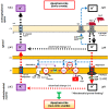
Schematic diagram illustrating the mechanism by which lactoferrin hypothetically induces an apoptosis-like process in yeast. Previously reported data on hLf-induced cell death have been incorporated into this model (2, 7, 22). In C. albicans cells, proton pumping through the Pma1p H+-ATPase (a) is essential for generation of a proton gradient (ΔpH) across the cytoplasmic membrane (CM) and for pH homeostasis, which is critical for cell survival. Under our experimental conditions, the blocking effect of hLf on Pma1p H+-ATPase (b) seems to induce a lethal perturbation in ionic homeostasis in two hypothetical coupled phases. Phase 1 is cytoplasmic ionic events, such as the intracellular accumulation of protons generated by an active metabolism due to the blocking effect of hLf on Pma1p H+-ATPase. To balance the electrical charge, accumulated protons pull potassium ions outside the cell through K+ channels. These previously reported ion-mediated events (2, 7, 22) will lead to phase 2, mitochondrial ionic events, where the previous cytoplasmic K+ efflux could facilitate a loss of mitochondrial potassium ions and the simultaneous reemplacement of mitochondrial potassium ions by protons entering via mtATPase, as suggested by incremental ATP synthesis. The coupled cytoplasmic and mitochondrial K+/H+ loops are indicated by the black and red thick arrows, respectively. Supporting this hypothesis, cellular protection was observed when the accumulation of protons in the mitochondrial matrix via mtATPase was prevented by (i) inhibition of the mtATPase with oligomycin and (ii) inhibition of proton translocation mediated by the respiratory chain, as suggested by the results obtained using piericidin A, anoxic conditions, or a respiration-deficient mutant. The hypothetical mitochondrial proton flooding process mediated by mtATPase could trigger the subsequent nonionic apoptotic events.

References

    1. García-Montoya IA, Cendón TS, Arévalo-Gallegos S, Rascón-Cruz Q. 2012. Lactoferrin a multiple bioactive protein: an overview. Biochim Biophys Acta 1820:226–236. doi:10.1016/j.bbagen.2011.06.018. - DOI - PMC - PubMed
    1. Andrés MT, Viejo-Díaz M, Fierro JF. 2008. Human lactoferrin induces apoptosis-like cell death in Candida albicans: critical role of K+-channel-mediated K+-efflux. Antimicrob Agents Chemother 52:4081–4088. doi:10.1128/AAC.01597-07. - DOI - PMC - PubMed
    1. Choi H, Lee W, Lee DG. 2013. A new concept on mechanism of antimicrobial peptides: apoptosis induction, p 775–785. In Ḿendez-Vilas A. (ed), Microbial pathogens and strategies for combating them: science, technology and education, vol 2 Formatex Research Center, Badajoz, Spain.
    1. Andrés MT, Fierro JF. 2010. Antimicrobial mechanism of action of transferrins: selective inhibition of H+-ATPase. Antimicrob Agents Chemother 54:4335–4342. doi:10.1128/AAC.01620-09. - DOI - PMC - PubMed
    1. Martínez-Muñoz GA, Kane P. 2008. Vacuolar and plasma membrane proton pumps collaborate to achieve cytosolic pH homeostasis in yeast. J Biol Chem 283:20309–20319. doi:10.1074/jbc.M710470200. - DOI - PMC - PubMed

MeSH terms

LinkOut - more resources