. 2016 May 3:17:81.
doi: 10.1186/s13059-016-0928-x.

Quantitative proteomics of the tobacco pollen tube secretome identifies novel pollen tube guidance proteins important for fertilization

Affiliations

Quantitative proteomics of the tobacco pollen tube secretome identifies novel pollen tube guidance proteins important for fertilization

Said Hafidh et al. Genome Biol. .

Abstract

Background: As in animals, cell-cell communication plays a pivotal role in male-female recognition during plant sexual reproduction. Prelaid peptides secreted from the female reproductive tissues guide pollen tubes towards ovules for fertilization. However, the elaborate mechanisms for this dialogue have remained elusive, particularly from the male perspective.

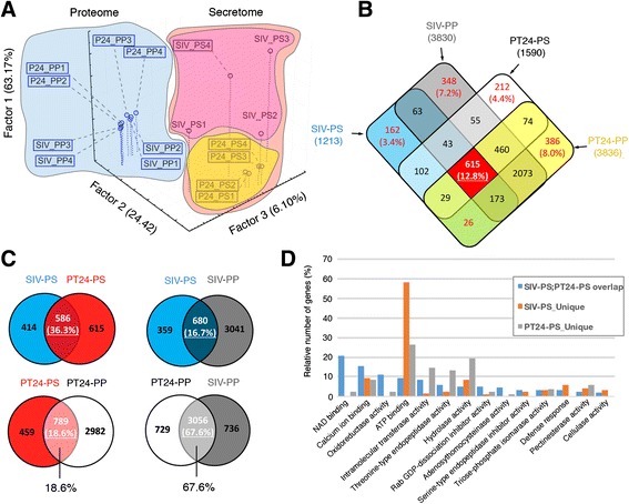
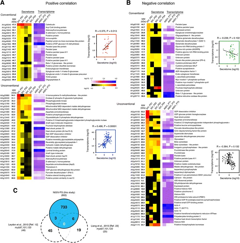
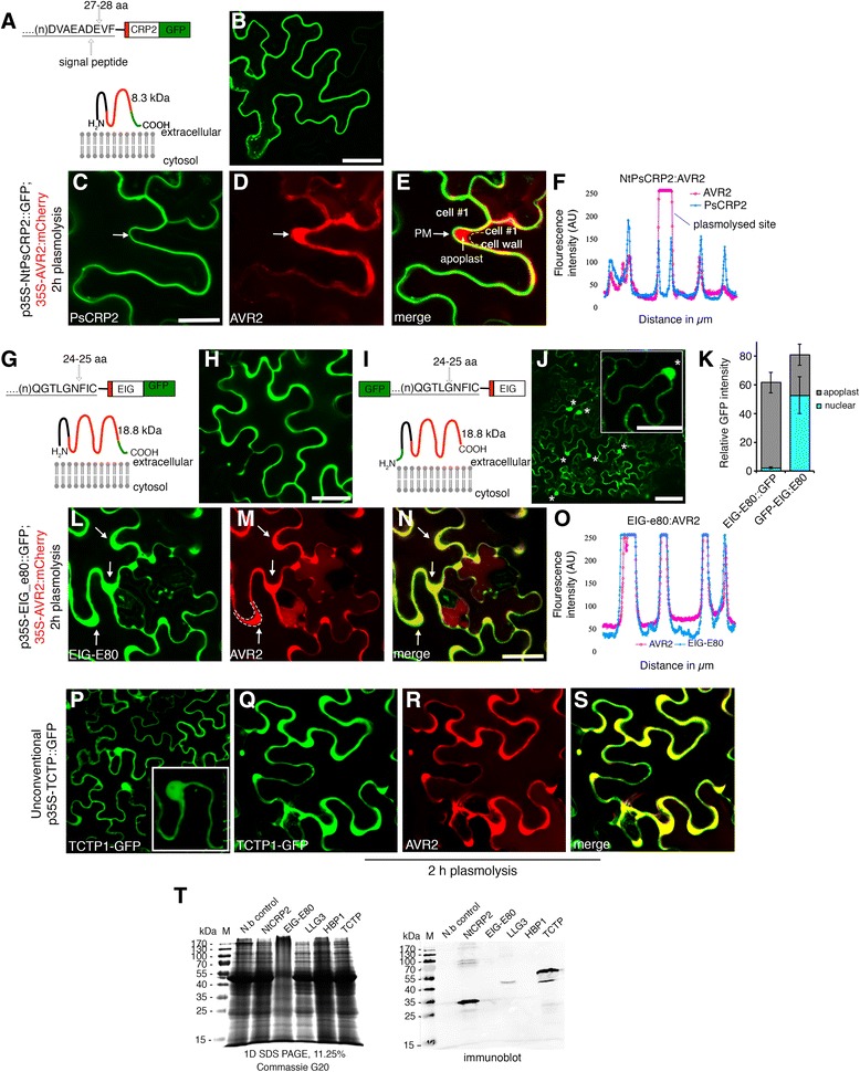
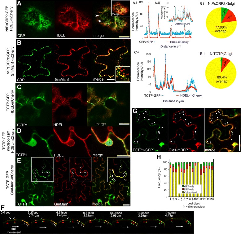
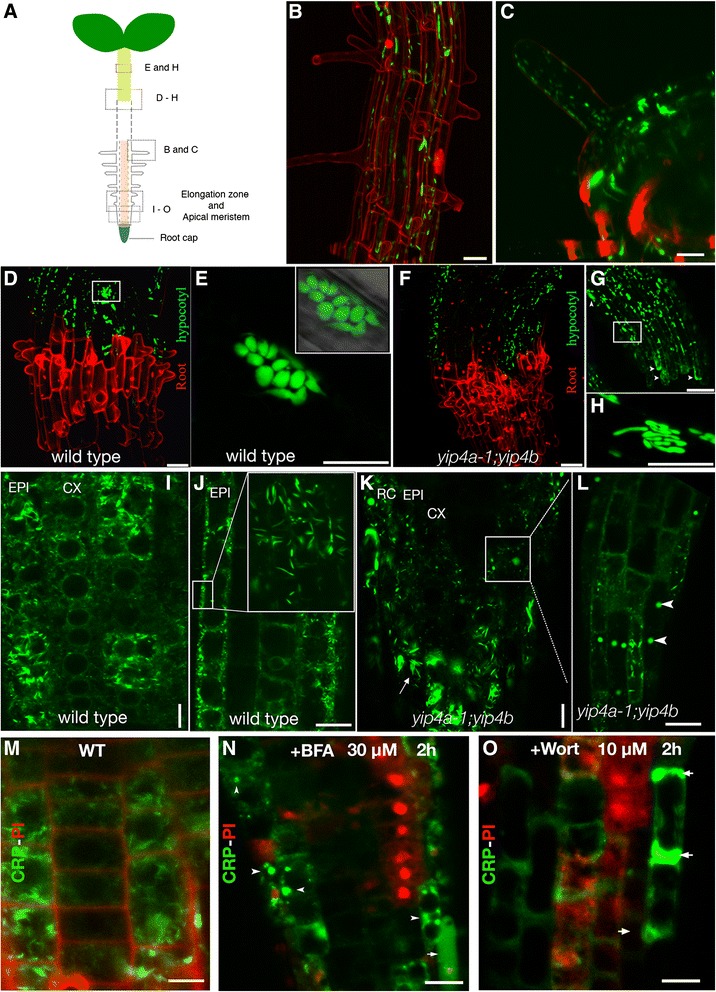
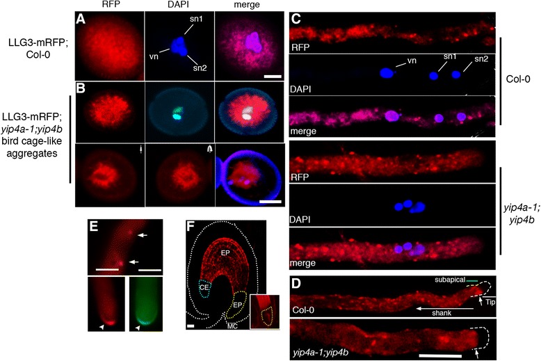
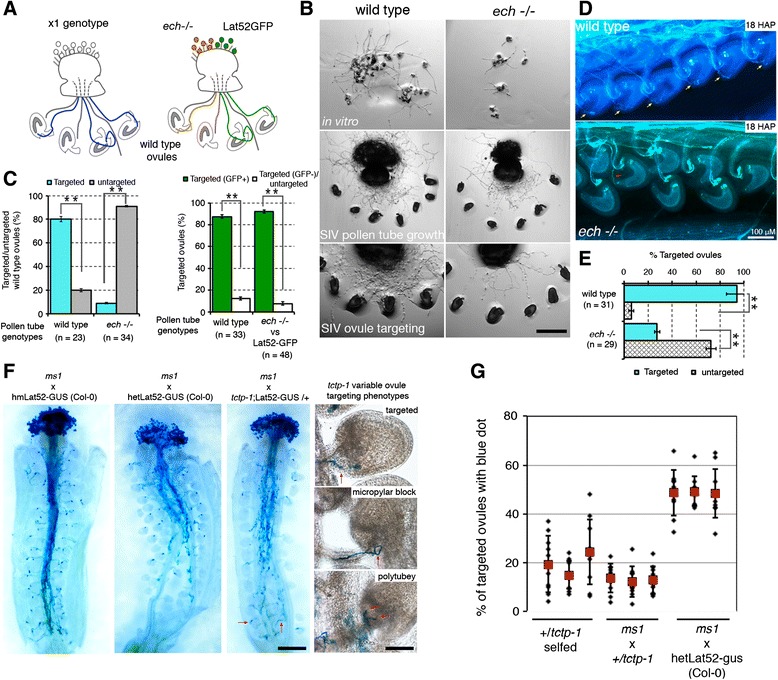
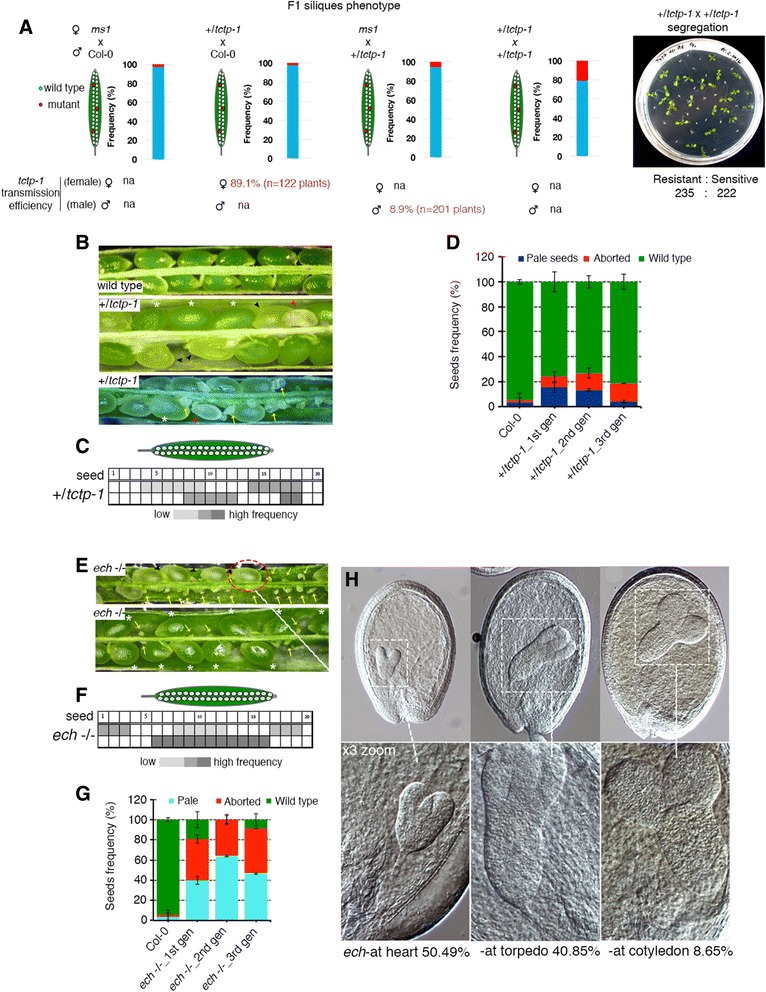
Results: We performed genome-wide quantitative liquid chromatography-tandem mass spectrometry analysis of a pistil-stimulated pollen tube secretome and identified 801 pollen tube-secreted proteins. Interestingly, in silico analysis reveals that the pollen tube secretome is dominated by proteins that are secreted unconventionally, representing 57 % of the total secretome. In support, we show that an unconventionally secreted protein, translationally controlled tumor protein, is secreted to the apoplast. Remarkably, we discovered that this protein could be secreted by infiltrating through the initial phases of the conventional secretory pathway and could reach the apoplast via exosomes, as demonstrated by co-localization with Oleisin1 exosome marker. We demonstrate that translationally controlled tumor protein-knockdown Arabidopsis thaliana plants produce pollen tubes that navigate poorly to the target ovule and that the mutant allele is poorly transmitted through the male. Further, we show that regulators of the endoplasmic reticulum-trans-Golgi network protein secretory pathway control secretion of Nicotiana tabacum Pollen tube-secreted cysteine-rich protein 2 and Lorelei-like GPI-anchor protein 3 and that a regulator of endoplasmic reticulum-trans-Golgi protein translocation is essential for pollen tube growth, pollen tube guidance and ovule-targeting competence.

Conclusions: This work, the first study on the pollen tube secretome, identifies novel genome-wide pollen tube-secreted proteins with potential functions in pollen tube guidance towards ovules for sexual reproduction. Functional analysis highlights a potential mechanism for unconventional secretion of pollen tube proteins and reveals likely regulators of conventional pollen tube protein secretion. The association of pollen tube-secreted proteins with marker proteins shown to be secreted via exosomes in other species suggests exosome secretion is a possible mechanism for cell-cell communication between the pollen tube and female reproductive cells.

Keywords: Cell-cell signaling; Double fertilization; Pollen tube guidance; Protein secretion.


Figures

Fig. 1
Fig. 1
Label-free, high-throughput LC-MS/MS quantification of the SIV pollen tube secretome. a Numbers of peptides identified by LC-MS/MS. Box plots summarize peptide number distribution per protein accession within a single sample replicate with the median value designated by the solid red line. Appended pie charts show the distribution of peptide counts in arbitrary bins. b SIV-PS samples showing the reproducibility and dynamics of the secreted protein groups (threefold or more abundant in at least two SIV-PS samples relative to unpollinated controls and identified by at least three or more peptides). c Size distribution of pollen tube-secreted proteins, showing predominant bias towards small secreted proteins. SIV-PS1–2 are used as representatives of all four replicates. d In silico analysis of pollen tube-secreted proteins using SMART and Pfam databases of overrepresented protein families and domains. The vertical white line indicates significance cutoff (p < 0.05). e Classified pollen tube-secreted proteins based on the Top3 algorithm showing the dominant presence of unconventionally secreted proteins, although these have comparable protein abundance to conventionally secreted proteins. f Heatmap derived from the Agilent tobacco microarray [19, 20] of transcripts encoding identified pollen tube-secreted proteins showing predominantly gametophytic enrichment. g Validated expression profile by semi-quantitative RT-PCR of selected pollen tube-secreted proteins assessed in this study. All samples analyzed were from N. tabacum. NtPsCRP1/2 Nicotiana tabacum cysteine-rich polypeptide protein 1 and 2, EIG-E80 elicitor inducible gene subE80, LLG3 Lorelei-like GPI-anchored protein 3, PT semi in vivo pollen tubes, Ov unfertilized ovules, Inf inflorescence
Fig. 2
Fig. 2
The secretome of pollen tubes grown through the pistil is unique from that of in vitro germinated pollen tubes and differs significantly from their respective proteomes. All sample types used for comparisons are representative of four replicates. a Three-dimensional principle component analysis of pollen tube secretomes (PS) and pollen tube proteomes (PP) from semi-in vivo (SIV) and 24 h in vitro germinated (PT24) pollen tubes. b Four-way Venn diagram of all sample types showing limited overlap between secretomes and proteomes but a relatively closer overlap between proteomes of SIV and in vitro germinated pollen tubes. c Pairwise comparison of sample similarities demonstrates that the secretome of pollen tubes grown through the pistil is unique from that of in vitro germinated pollen tubes and the pollen tube secretome is different from their respective pollen tube total proteomes, suggesting that regulated exo- and endocytosis occurs during pollen tube protein secretion. d GO term comparison of SIV and in vitro pollen tube secretomes, highlighting enriched activities where they overlap and protein accessions unique between the two sample types
Fig. 3
Fig. 3
Semi-in vivo transcriptome–secretome correlation. Pollen tube-secreted proteins showed moderate positive (a) and negative (b) correlation, but not linear correlation, with transcripts of the Arabidopsis SIV pollen tube transcriptome [15]. To the right of each heatmap are scatter plots with Pearson correlation coefficient scores and confidence values at p< 0.01. c Comparison of tobacco SIV-PS with downstream targets of MYB97, 101, and 120 identified in [12, 23]
Fig. 4
Fig. 4
Interference of the N-terminal signal peptide compromises apoplastic localization of the EIG-E80 pollen tube-secreted protein. Secretion of pollen tube-secreted proteins to the proximity of the plasma membrane and the apoplast of tobacco leaf epidermal cells is signal peptide-dependent. a, b Chimeric construct of pollen tube-secreted cysteine-rich polypeptide protein 2 (NtPsCRP2) and its predicted topology showing extracellular localization. c–e Verification of NtPsCRP2 localization near the plasma membrane (PM) by plasmolysis of tobacco epidermal cells co-infiltrated with apoplastic viral AVR2-mCherry. The arrow shows plasmolyzed regions with detached plasma membrane and apoplastic localized PR1a-AVR2-mCherry. f Two-channel confocal laser scan profile during co-localization of NtPsCRP2 with apoplastic marker AVR2-mCherry following plasmolysis. AU arbitrary units. g, h Chimeric construct of elicitor-induced protein E80 (EIG-E80) and its predicted topology showing apoplastic/extracellular localization. i, j Reconstituted GFP:EIG-E80 chimeric construct with blocked signal peptide resulted in partial apoplastic localization and predominant nuclear localization instead. k Subcellular quantification of the modified GFP:EIG-E80 expression. Error bars represent ± standard deviation. l–o Verification of EIG-E80:GFP apoplastic localization by plasmolysis with viral AVR2 apoplastic marker. Arrows indicate plasmolysed regions. p Localization of the unconventionally secreted tobacco pollen tube protein Translationally controlled tumor protein (TCTP) showing nucleoplasm, cytosol, and apoplastic localization. q–s Verification of TCTP apoplastic localization. t Immunodetection verification of ectopically expressed GFP chimeric pollen tube-secreted proteins with rat anti-GFP monoclonal antibodies in tobacco epidermal cells. Commassie G250 stain of total protein extract (left) and immunoblot (right) 48 h post-infiltration. Scale bars = 10 μM. N.b untransformed Nicotiana benthamiana
Fig. 5
Fig. 5
NtPsCRP2 and unconventionally secreted NtTCTP enter the ER and co-localize with the Golgi marker GmMan1 and exosome marker Ole1. a Confocal micrographs showing co-localization NtPsCRP2-GFP with the ER retention marker HDEL-mCherry with the exception of GFP foci (white arrowheads). Two-channel profile overlap (a-i). Emphasis on the lack of complete overlap (black arrowheads) of ER-HDEL with NtPsCRP2-GFP foci (a-ii). b NtPsCRP2 co-localization with the ER–Golgi vesicle marker GmMan1-mCherry from soybean. Frequency of co-localization (29 confocal slices, n = 340) (b-i). c Co-localization of unconventionally secreted NtTCTP with ER HDEL-mCherry. Visualization of ER co-localization of NtTCTP by two-channel pixel-intensity profiling (c-i). d NtTCTP also co-localizes with the ER–nuclear lamina and is present in the nucleoplasm. e NtTCTP co-localization with the Golgi vesicle marker GmMan1-mCherry. Arrowheads indicate granules formed by TCTP-GFP and those by GmMan1-mCherry. e–i Frequency of overlapping foci observed in 32 confocal slices (n = 292). f Dynamics of NtPsCRP2-GFP foci (green) and Golgi-derived vesicles (red) in the proximity of the plasma membrane. The top white arrows point to tethered NtPsCRP2-GFP foci over time, the red arrowheads Golgi vesicles alone, the green arrowheads NtPsCRP2-GFP foci alone, and the red-green arrowheads co-localized NtCRP1-Golgi signals, and foci 1–4 are additional NtCRP1-Golgi co-localized vesicles migrating clockwise towards the membrane horizon over time. Note the appearance and disappearance of co-localized NtPsCRP2-Golgi vesicles as well as NtPsCRP2-GFP foci alone. Scale bars = 20 μM. g NtTCTP co-localized with Ole1 in potential nanovesicle exosomes. Co-expression of NtTCTP-GFP with AtOle1-mRFP in tobacco leaf epidermal cells revealed granular co-localizations (marked with arrowheads). h Frequency of co-localizations observed from multiple leaf discs. Scale bars = 10 μM, RFP red fluorescent protein
Fig. 6
Fig. 6
Simultaneous knockdown of YIP4A/B perturbed NtPsCRP2 secretion in seedlings. a An Arabidopsis seedling showing regions analyzed in this study. b, c NtPsCRP2-GFP localized less frequently in young root hairs but more frequently in mature root hairs, both in the wild type and in the yip4a-1;yip4b double mutant. d NtPsCRP2 secretion in 4-day-old etiolated wild-type seedlings showing localization in epidermal cells of the hypocotyl. The secretion was restricted at the hypocotyl–root junction. Propidium iodide (red) marks the beginning of the root. e Close-up of NtPsCRP2-GFP in elongated epidermal cells of the hypocotyl showing accumulation in spherical, aggregated endosomal-like vesicles. f, g Conversely, in yip4a-1;yip4b seedlings, NtPsCRP2 secretion was severely distorted, showing diffuse localization and non-uniform protein aggregates (arrowheads). h The spherical marked endosomal aggregates observed in the wild type were substituted with rod-like labeled organelles in yip4a-1;yip4b hypocotyl. i NtPsCRP2 localization at the root tip of wild-type seedlings marking secretory organelles. EPI epidermis, CX cortex. j Magnified root epidermal cells from wild type showing NtPsCRP2-marked organelles constrained to the vicinity of the plasma membrane (white rectangle) and also localized in ER-body-like organelles (inset). k, l In yip4a-1;yip4b, NtPsCRP2-GFP localization in secretory organelles at the root apical meristem appeared largely disorganized, showing severe organelle aggregations resembling BFA-like compartments (arrowheads). The ER-body-like localization (arrow) was maintained, suggesting no effect on ER-body biogenesis in yip4a-1;yip4b mutants. m–o Perturbed secretion of NtPsCRP2-GFP was also recapitulated following BFA or wortmannin treatment. Arrowheads show NtPsCRP2-GFP BFA compartments, arrows point to diffusely aggregated NtPsCRP2-GFP aberrant secretion. RC lateral root cap PI propidium iodide stain, WT wild type. Scale bars = 10 μM
Fig. 7
Fig. 7
LLG3 secretion is compromised in yip4a-1;yip4b mutant pollen and pollen tubes. a Subcellular localization of LLG3-mRFP under native promoter showing cytosolic foci-like aggregates. vn vegetative cell nucleus, sn sperm cell nuclei. b Top: in the yip4a-1;yip4b mutant, the majority of the pollen grains displayed distinct cytosolic aggregates different from those observed in the wild type. Bottom: z-stack projection of 25 confocal slices showing distinct vegetative cell “bird cage-like” localization that was not observed in wild-type pollen grains. c Localization of LLG3-mRFP in wild-type pollen tubes grown in vitro for 16 h showing localization in likely secretory vesicles of uniform size and partially in the pollen tube ER (top three panels). In contrast, yip4a-1;yip4b mutant pollen tubes displayed a significantly higher frequency of LLG3-mRFP-marked endomembrane aggregated vesicles of variable sizes (bottom three panels). However, the ER localization was not greatly affected. d LLG3-mRFP also specifically accumulated at the subapical domain (top panel, arrow). This accumulation was also not significantly affected in yip4a-1;yip4b mutant pollen tubes (bottom panel). e In tobacco pollen tubes, similar localization in secretory vesicles was also observed (top panel, arrows) and occasionally LLG3-mRFP as well as LLG3-sGFP showed pollen tube tip-specific localization (bottom panels, arrowheads). f Absence of LLG3-mRFP protein in Arabidopsis unfertilized ovules and embryos soon after fertilization (18 h after pollination). Rarely, LLG3-mRFP could be detected at the embryo proper (EP) zone in some fertilized ovules. MC micropylar, EP endosperm proper, CE chalaza end endosperm. Scale bars for a and b = 30 μM and for c–e = 10 μM
Fig. 8
Fig. 8
TCTP- and ECHIDNA-deficient pollen tubes consistently fail to target ovules. a The SIV ovule-targeting assay. Pollen tube targeting competence was assessed using a single pollen genotype or in a one-to-one genotype competition assay. b ech-/- pollen grains showed reduced but adequate pollen tube germination in vitro as well as semi-in vivo but showed significantly reduced ovule-targeting competence semi-in vivo. c Quantification of pollen tube ovule-targeting competence semi-in vivo of ech-/- plants in a single genotype or mixed genotype competition assay. n is the number of pistil explants assessed, asterisks denote significant differences assessed with Student’s t-test (p < 0.01). d Aniline blue stain of wild-type and ech-/- pollinated wild-type pistils 18 h after pollination (HAP) showing near complete lack of ovule targeting in ech-/- pollinated pistils (red arrow) compared with wild type-pollinated pistils (yellow arrows), verifying the semi-in vivo observations in c. e Frequency of ovule targeting by pollen tubes in aniline blue-stained pistils. All score variances (asterisk) are statistically significant (Student’s t-test, p < 0.01). Error bars represent standard deviation (stdv). f Blue dot assay by GUS-staining of ms1 pistils pollinated with wild-type pollen grains homozygous or heterozygous for Lat52-GUS and Lat52-GUS;tctp-1/+ 18 HAP. Red arrows point to tctp-1 mutant pollen tubes targeting ovules at the bottom of the pistil, suggesting tctp-1 pollen tube growth is not greatly impaired. Insets: variable mistargeting phenotypes observed in tctp-1/+ pollinated pistils. g Counts of “blue dots” revealed ovule targeting was greatly impaired in tctp-deficient pollen tubes
Fig. 9
Fig. 9
Loss of TCTP and ECHIDNA functions also impairs post-fertilization events. a Reciprocal test crosses of the tctp-1 mutation showing low penetrance male-specific phenotypic induction in F1 siliques as well as significantly reduced male transmission efficiency scored by PCR genotyping as well as GUS-staining of LAT52-GUS-tagged T-DNA insertion. The micrograph on the far right shows seedling segregation of self-fertilized +/​tctp-1 plants, supporting the near complete block of tctp-1 allele transmission through the male. b Dissected siliques of wild-type and self-fertilized +/​tctp-1 plants. Asterisks indicate wild-type-like seeds, black arrowheads indicate underdeveloped chlorotic mutant seeds, red arrowheads and yellow arrows show arrested embryos with failed embryogenesis soon after fertilization as well as unfertilized ovules. c Frequency and positioning of mutant seeds within dissected siliques. The random distribution of mutant seeds within +/​tctp-1 siliques suggests tctp-1 mutant pollen tube growth is competent. d Frequency of tctp-1 mutant seed phenotypic classes observed (n = 25 siliques per line). Error bars represent standard deviation (stdv). e Dissected siliques of self-fertilized ech-/- plants showing the high frequency of failed fertilization, defective embryogenesis, as well as “wild-type-like” seeds (asterisks). f Frequency and random positioning of ech mutant embryos/unfertilized ovules within ech-/- self-fertilized siliques implying ech pollen tubes can grow to the base of the pistil and target ovules for fertilization. g Frequency of mutant seed phenotypic classes observed (n = 25 siliques per line). Error bars represent standard deviation (stdv). h Embryogenesis lethality in ECHIDNA-deficient embryos showing stages and frequency of embryo arrest. Scale bars = 10 μM. gn germ cell nuclei, na not applicable, sn sperm cell nuclei, vn vegetative cell nuclei, wt wild type

Comment in

References

    1. Maheshwari P. An introduction to embryology of angiosperms. New York: McGraw-Hill; 1950.
    1. Yadegari R, Drews GN. Female gametophyte development. Plant Cell. 2004;16:S133–41. doi: 10.1105/tpc.018192. - DOI - PMC - PubMed
    1. Okuda S, Tsutsui H, Shiina K, Sprunck S, Takeuchi H, Yui R, et al. Defensin-like polypeptide LUREs are pollen tube attractants secreted from synergid cells. Nature. 2009;458:357–61. doi: 10.1038/nature07882. - DOI - PubMed
    1. Takeuchi H, Higashiyama T. Attraction of tip-growing pollen tubes by the female gametophyte. Curr Opin Plant Biol. 2011;14:614–21. doi: 10.1016/j.pbi.2011.07.010. - DOI - PubMed
    1. Marton ML, Fastner A, Uebler S, Dresselhaus T. Overcoming hybridization barriers by the secretion of the maize pollen tube attractant ZmEA1 from Arabidopsis ovules. Curr Biol. 2012;22:1194–8. doi: 10.1016/j.cub.2012.04.061. - DOI - PubMed

Publication types

LinkOut - more resources