. 2016 Aug;42(8):1890-902.
doi: 10.1016/j.ultrasmedbio.2016.03.018. Epub 2016 Apr 29.

Non-Invasive Ultrasound Liver Ablation Using Histotripsy: Chronic Study in an In Vivo Rodent Model

Affiliations

Non-Invasive Ultrasound Liver Ablation Using Histotripsy: Chronic Study in an In Vivo Rodent Model

Eli Vlaisavljevich et al. Ultrasound Med Biol. 2016 Aug.

Abstract

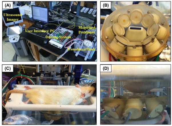
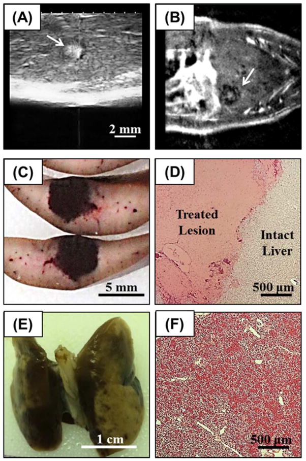
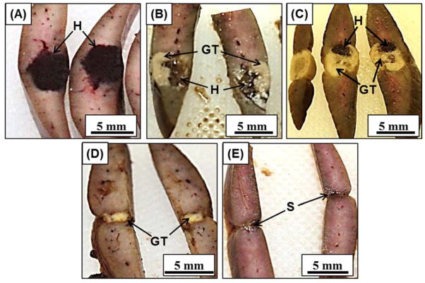
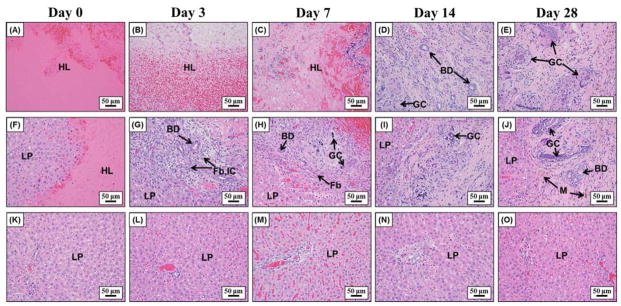
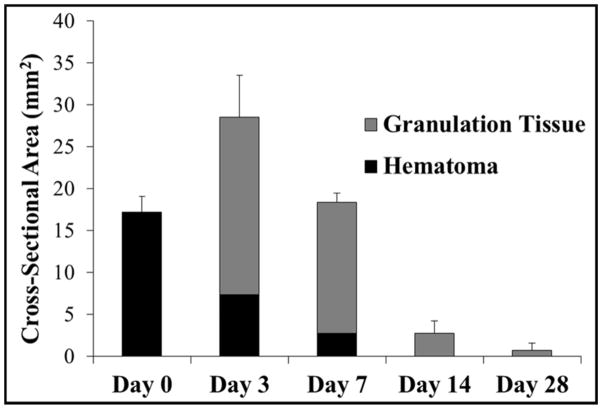
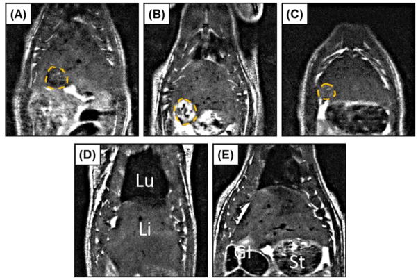
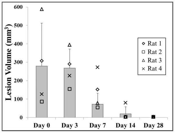
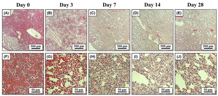
Hepatocellular carcinoma, or liver cancer, has the fastest growing incidence among cancers in the United States. Current liver ablation methods are thermal-based and share limitations due to the heat sink effect from the blood flow through the highly vascular liver. Recently, our group has investigated histotripsy as a non-invasive liver cancer ablation method. Histotripsy is a non-thermal ultrasonic ablation method that fractionates tissue through the control of acoustic cavitation. Previous experiments in an in vivo porcine model show that histotripsy can create well-confined lesions in the liver through ribcage obstruction without damaging the overlying ribs and other tissues. Histotripsy can also completely fractionate liver tissue surrounding major vessels while preserving the vessels. In this study, we investigate the long-term effects of histotripsy liver ablation in a rodent model. We hypothesize that the fractionated histotripsy lesion will be resorbed by the liver, resulting in effective tissue healing. To test this hypothesis, the livers of 20 healthy rats were treated with histotripsy using an 8-element 1-MHz histotripsy transducer. Rats were euthanized after 0, 3, 7, 14 and 28 days (n = 4). In vivo and post mortem results showed histotripsy lesions were successfully generated through the intact abdomen in all 20 rats. Magnetic resonance imaging found primarily negative contrast on day 0, positive contrast on day 3 and rapid normalization of signal intensity thereafter (i.e., signal amplitude returned to baseline levels seen in healthy liver tissue). Histologically, lesions were completely fractionated into an acellular homogenate. The lesions had a maximum cross-sectional area of 17.2 ± 1.9 mm(2) and sharp boundaries between the lesion and the healthy surrounding tissue after treatment. As the animals recovered after treatment, the histotripsy tissue homogenate was almost completely replaced by regenerated liver parenchyma, resulting in a small fibrous lesion (<1 mm(2) maximum cross-section) remaining after 28 d. The results of this study suggest that histotripsy has potential as a non-invasive liver ablation method for effective tissue removal.

Keywords: Cavitation; Histotripsy; Liver cancer; Non-invasive tissue ablation; Ultrasound.


Figures

Figure 1
Figure 1. Histotripsy in vivo rat liver ablation experimental setup
(A) A 1 MHz histotripsy therapy transducer with coaxially aligned ultrasound imaging probe was attached to a motorized 3D positioning system controlled using a PC console. (B) The histotripsy transducer consisted of 8 transducer elements in a ring with the imaging probe inserted in the center. (C) For treatment, an anesthetized rat was placed on a stage above the histotripsy transducer. (D) The transducer was placed inside a tank of degassed water beneath the anesthetized rat, and histotripsy was noninvasively applied to the liver through the intact abdomen.
Figure 2
Figure 2. Acoustic waveform
Image shows an example acoustic waveform generated by the 8-element 1 MHz histotripsy small animal transducer and measured by the FOPH.
Figure 3
Figure 3
(A) Histotripsy treatments were applied to the rat liver guided by real-time ultrasound imaging (arrow indicates histotripsy bubble cloud). After treatment, liver lesions were assessed using (B) MRI (lesion indicated by arrow), followed by assessment with (C) gross morphology and (D) histology. In addition to the liver, the inferior lobes of the lungs were harvested after treatment and assessed using (E) gross morphology and (F) histology.
Figure 4
Figure 4
Histotripsy liver lesions were assessed for gross morphology after animals were sacrificed on days 0, 3, 7, 14, 28 (AE). Darkly colored hematomas seen on (A) day 0 were replaced with granulation tissue on (B) day 3 and (C) day 7. The granulation tissue was mostly replaced by regenerated liver parenchyma on (D) day 14 and (E) day 28, leaving a small contracted scar. H=hematoma, GT=granulation tissue, S=scar
Figure 5
Figure 5
Representative histology images showing the center of the lesion (top row), the lesion interface (middle row), and the surrounding liver parenchyma (bottom row) after day 0, 3, 7, 14, and 28. Initially, lesions were almost solely composed of extravasated red blood cells and fibrin on day 0 (A, F). Fibroblasts started to grow into the lesion from the interface together with inflammatory cells and foci of bile ductular proliferation at day 3 (G) and day 7 (H). At day 14 (D, I) and day 28 (E, J), the majority of the lesion was replaced by regenerated liver parenchyma with only a small focus of residual scar and foreign body giant cells with calcified undigested debris in their cytoplasm. The surrounding liver parenchyma is unremarkable with no significant inflammation, necrosis, or fibrosis (K–O). LP=liver parenchyma, HL=histotripsy lesion, BD=bile ducts, Fb=fibroblasts, IC= inflammatory cells, M=macrophages, GC=giant cells.
Figure 6
Figure 6
Plot shows the average maximum cross-sectional area of the histotripsy liver lesions measured over the course of the experiment. The area of the hematoma and granulation tissue components of the lesion, as well as the total lesion cross-sectional area, were compared for rats euthanized after day 0, 3, 7, 14, and 28. Following an increase in the maximum cross-sectional area between day 0 and 3, there was a rapid reduction in lesion size over the remainder of the study. All differences in lesion size were significant (p<0.05).
Figure 7
Figure 7
Representative MRI images for one rat acquired on days 0, 3, 7, 14, and 28 (A–E), illustrating approximately the maximum cross-sectional area of the histotripsy lesion (Lesion=dashed orange line; Lu=lungs, Li=liver, St=stomach, GI=gastrointestinal tract). Lesions showed primarily negative contrast on day 0 (A) and positive contrast on day 3 (B). Signal intensity rapidly normalized at later time points (C–E), with no lesion quantifiable in any animal by day 28 (E).
Figure 8
Figure 8
Plot shows the lesion volume measured by MRI for rats euthanized after day 28 (n=4). No significant difference in lesion size was observed between day 0 and day 3 (p>0.05). After day 3, there was a significant decrease in lesion volume over the remainder of the study (p<0.05) with no quantifiable lesions observed after day 28.
Figure 9
Figure 9. Lung Histology
Histology images of the inferior lobes of the lung after days 0, 3, 7, 14, 28 using 4x (A–E) and 40x magnification (F–J). Results showed localized lung hemorrhage on day 0 (A, F), indicated by the presence of red blood cells throughout the alveolar space. After recovery from therapy, the number of red blood cells inside the alveolar space steadily decreased, with minimal hemorrhage remaining after day 28 (E, J).

References

    1. ACS. Cancer Facts and Figures 2015. American Cancer Society; 2015.
    1. Alkins R, Huang Y, Pajek D, Hynynen K. Cavitation-based third ventriculostomy using MRI-guided focused ultrasound. J Neurosurg. 2013;119:1520–9. - PMC - PubMed
    1. Allen SP, Hall TL, Cain CA, Hernandez-Garcia L. Controlling cavitation-based image contrast in focused ultrasound histotripsy surgery. Magn Reson Med. 2014 - PubMed
    1. Apfel RE, Holland CK. Gauging the likelihood of cavitation from short-pulse, low-duty cycle diagnostic ultrasound. Ultrasound Med Biol. 1991;17:179–85. - PubMed
    1. Aschoff AJ, Merkle EM, Wong V, Zhang Q, Mendez MM, Duerk JL, Lewin JS. How does alteration of hepatic blood flow affect liver perfusion and radiofrequency-induced thermal lesion size in rabbit liver? J Magn Reson Imaging. 2001;13:57–63. - PubMed