Review
. 2016 Jul;12(7):426-39.
doi: 10.1038/nrneph.2016.54. Epub 2016 May 3.

Developmental signalling pathways in renal fibrosis: the roles of Notch, Wnt and Hedgehog

Affiliations
Review

Developmental signalling pathways in renal fibrosis: the roles of Notch, Wnt and Hedgehog

Maria Edeling et al. Nat Rev Nephrol. 2016 Jul.

Abstract

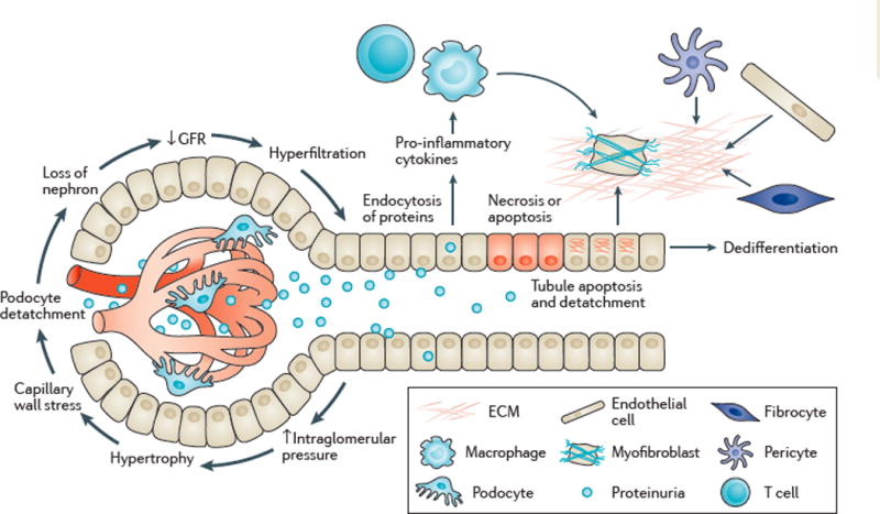
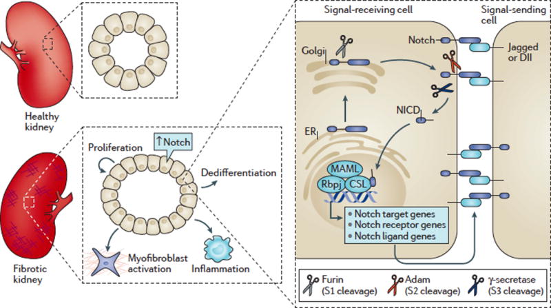
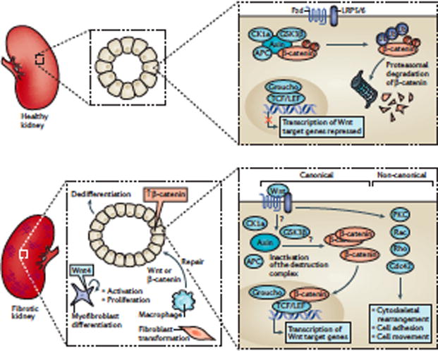
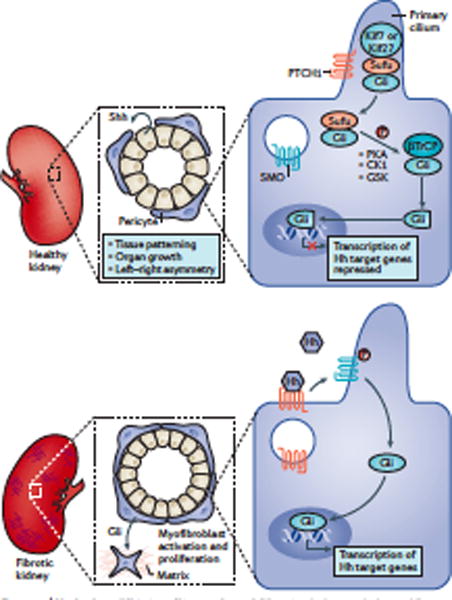
Kidney fibrosis is a common histological manifestation of functional decline in the kidney. Fibrosis is a reactive process that develops in response to excessive epithelial injury and inflammation, leading to myofibroblast activation and an accumulation of extracellular matrix. Here, we describe how three key developmental signalling pathways - Notch, Wnt and Hedgehog (Hh) - are reactivated in response to kidney injury and contribute to the fibrotic response. Although transient activation of these pathways is needed for repair of injured tissue, their sustained activation is thought to promote fibrosis. Excessive Wnt and Notch expression prohibit epithelial differentiation, whereas increased Wnt and Hh expression induce fibroblast proliferation and myofibroblastic transdifferentiation. Notch, Wnt and Hh are fundamentally different signalling pathways, but their choreographed activation seems to be just as important for fibrosis as it is for embryonic kidney development. Decreasing the activity of Notch, Wnt or Hh signalling could potentially provide a new therapeutic strategy to ameliorate the development of fibrosis in chronic kidney disease.


Conflict of interest statement

Competing interests

The Susztaklab received research support from Boehringer Ingelheim, Biogen and Lilly.

Figures

Figure 1
Figure 1. Development of kidney fibrosis
Glomerular hyperfiltration is associated with glomerular hypertrophy, which results in podocyte loss. Loss of podocytes causes albuminuria and glomerulosclerosis, and nephron loss. Hyperglycaemia, increased levels of fatty acids and proteinuria cause epithelial damage including apoptosis, detachment and dedifferentiation of epithelial cells, and increased expression of pro-inflammatory cytokines. Increased cytokine expression causes increased macrophage, T-cell, and mast-cell influx. Epithelial damage also induces myofibroblast transformation and production of extracellular matrix (ECM). GFR, glomerular filtration rate.
Figure 2
Figure 2. Notch signalling in renal fibrosis
In healthy adult kidneys, the activity of Notch signaling is low. Acute and chronic kidney disease is associated with increased epithelial expression of Notch signaling memebers. The Notch receptor is expressed on signal-receiving cells. Binding of the Notch ligand on the signal-sending cell to the receptor results in Notch cleavage (by the ƴ-secretase complex) and release of the Notch intracellular domain (NICD). In the nucleus, NICD binds to other transcriptional regulators such as Rbpj, and MAML for regulating gene expression. In chronic kidney disease, increased Notch activity can be observed in renal epithelial cells. Increased Notch receptor expression is associated with epithelial dedifferentiation, myofibroblast activation, matrix deposition, and the inflammatory response.
Figure 3
Figure 3. Dysregulation of Wnt signalling leads to the development of renal fibrosis
In the absence of the Wnt ligands (inactive Wnt signaling), cytoplasmic β-catenin is phosphorylated by the ‘destruction complex’ containing the glycogen synthase kinase 3b (GSK3b), the scaffolding protein axin, casein kinase 1a (CK1a), and the adenomatous polyposis coli protein (APC), which is followed by ubiquitin-mediated proteasomal degradation. Binding of Wnt to Frizzled (Fzd) and LRP5/6 receptor proteins inactivates GSK3b and disassembles the ‘destruction complex’. Accumulation of non-phosphorylated β-catenin in the cytoplasm leads to its translocation into the nucleus (canonical Wnt signaling). There, β-catenin replaces Groucho from TCF/LEF and activates Wnt target gene expression. Increased tubule-specific β-catenin expression has been described in chronic kidney disease (CKD). Myofibroblasts can express Wnt4, which induces proliferation of resident fibroblasts and their transformation into myofibroblasts. Wnt/β-catenin signalling in pericytes and interstitial fibroblasts exhibits spontaneous myofibroblast differentiation, the relevant step in fibrosis development. Macrophage expression of Wnt7b is associated with epithelial repair. The ß-catenin-independant (non-canonical) Wnt signaling pathway regulates cytoskeleton rearrangement, cell adhesion and cell movement via the kinases Rho, Rac and Cdc42.
Figure 4
Figure 4. Hedgehog (Hh) signalling and renal fibrosis
In humans, Hh molecules bind to the receptor Patched1 (PTCH1), which leads to localization of SMO to the primary cilium. Ciliary SMO mediates the translocation of full-length GLI to the nucleus. GLI binds to DNA and activates expression of Hh target genes. In the steady state of Hh signaling GLI proteins are sequestered by Kif7/Kif27, Fused (Fu) and the Suppressor of Fused (Sufu) followed by phosphorylation by protein kinase A (PKA), casein kinase 1 (CK1) and glycogen synthase kinase 3b (GSK3b). This modification allows proteolytical cleavage of GLI by b-transducin repeat containing protein (bTrCP). The now C-terminal shortened GLI repressor binds to the DNA for inhibiting Hh target gene transcription Hh ligands are expressed in tubular epithelial cells, and interstitial cells respond to Hh ligands. Gli1-expressing pericytes have an important role in interstitial fibrosis, undergoing myofibroblast transformation and proliferation.

References

    1. Jha V. Chronic kidney disease: global dimension and perspectives. Lancet. 2013;382:260–272. doi: 10.1016/S0140-6736(13)60687-X. - DOI - PubMed
    1. Liu Y. Renal fibrosis: new insights into the pathogenesis and therapeutics. Kidney Int. 2006;69:213–217. doi: 10.1038/sj.ki.5000054. - DOI - PubMed
    1. Tonelli M, et al. Chronic kidney disease and mortality risk: a systematic review. J Am Soc Nephrol. 2006;17:2034–2047. doi: 10.1681/ASN.2005101085. - DOI - PubMed
    1. Reidy K, Kang HM, Hostetter T, Susztak K. Molecular mechanisms of diabetic kidney disease. J Clin Invest. 2014;124:2333–2340. doi: 10.1172/JCI72271. - DOI - PMC - PubMed
    1. Duffield JS. Cellular and molecular mechanisms in kidney fibrosis. J Clin Invest. 2014;124:2299–2306. doi: 10.1172/JCI72267. - DOI - PMC - PubMed

Publication types