Review
. 2016 May 2;8(5):a019372.
doi: 10.1101/cshperspect.a019372.

Maintenance of Epigenetic Information

Affiliations
Review

Maintenance of Epigenetic Information

Geneviève Almouzni et al. Cold Spring Harb Perspect Biol. .

Abstract

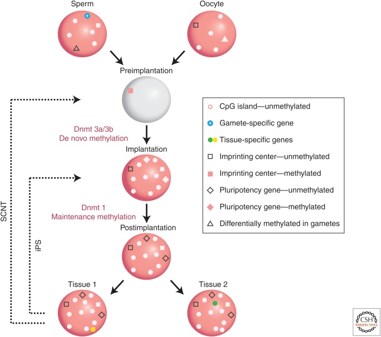
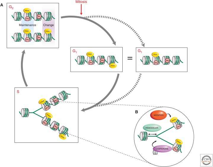
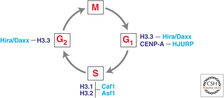
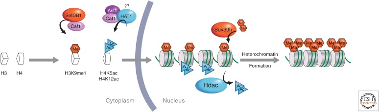
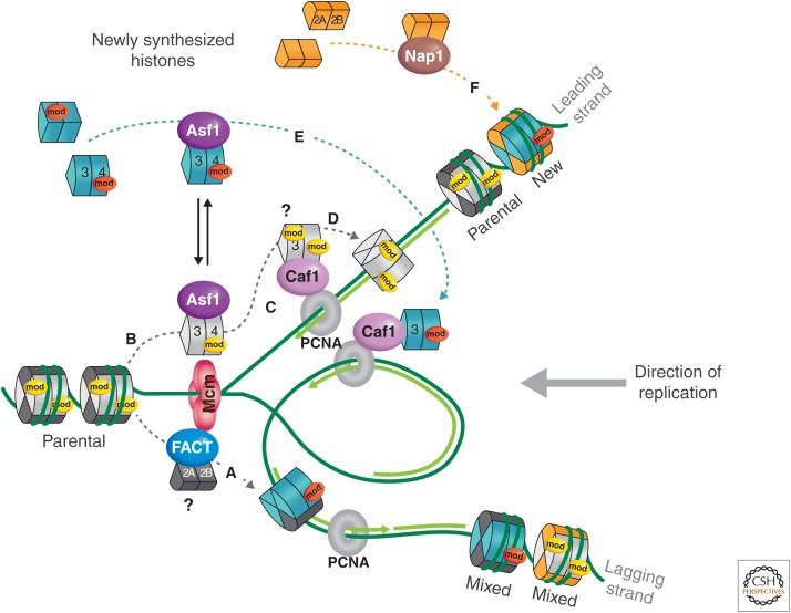
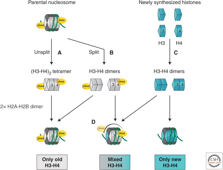
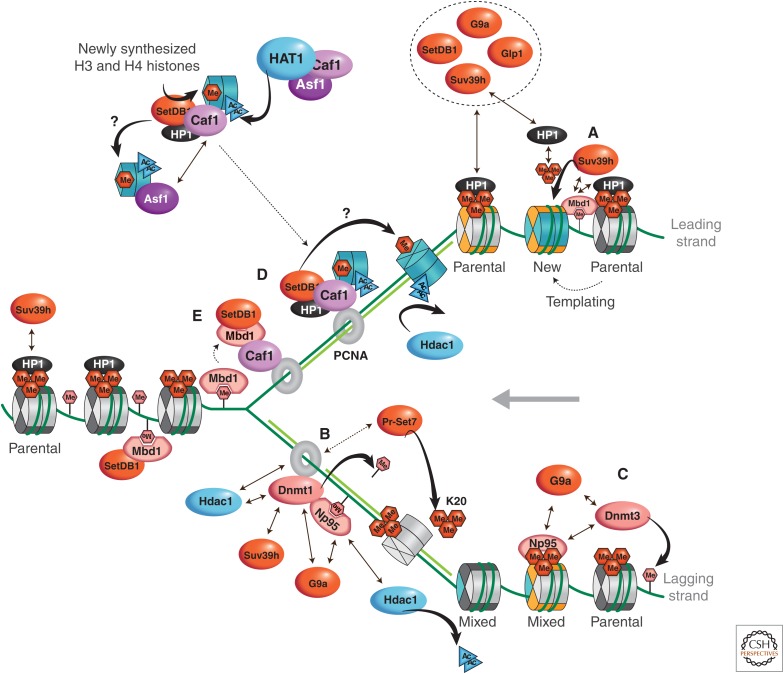
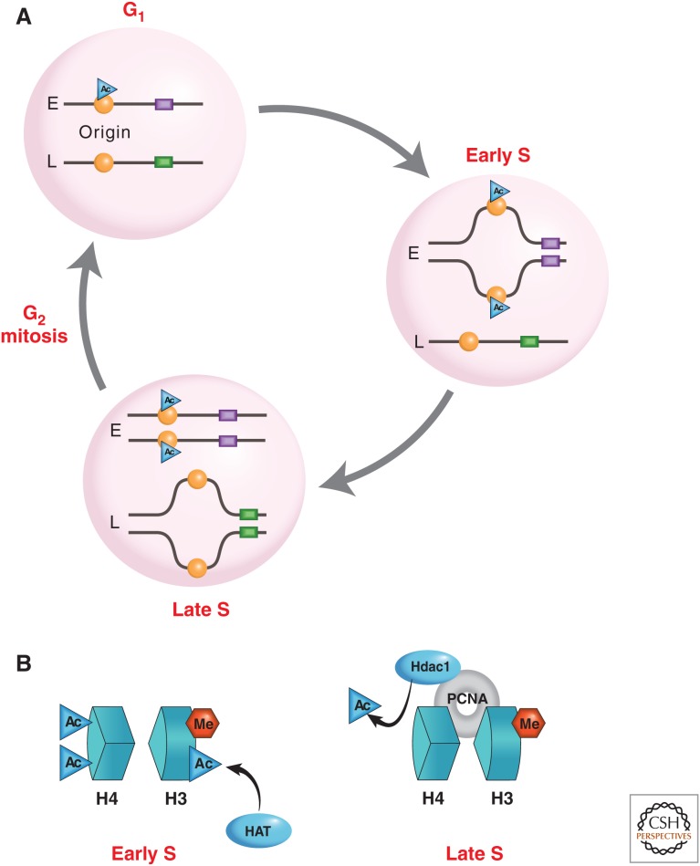
The genome is subject to a diverse array of epigenetic modifications from DNA methylation to histone posttranslational changes. Many of these marks are somatically stable through cell division. This article focuses on our knowledge of the mechanisms governing the inheritance of epigenetic marks, particularly, repressive ones, when the DNA and chromatin template are duplicated in S phase. This involves the action of histone chaperones, nucleosome-remodeling enzymes, histone and DNA methylation binding proteins, and chromatin-modifying enzymes. Last, the timing of DNA replication is discussed, including the question of whether this constitutes an epigenetic mark that facilitates the propagation of epigenetic marks.


Figures

Figure 1.
Figure 1.
Generation of DNA methylation patterns during development. Gametic cells have a bimodal pattern of methylation with most regions methylated and CpG islands unmethylated (gray circles). Imprinting centers are methylated in one gamete (pink square), but not the other (white square). Gamete-specific genes (blue ring) are unmethylated. Some genes (triangles) are specifically unmethylated (gray) in one gamete. Almost all methylation in the gametes is erased (gray) in the preimplantation embryo, but imprinting centers retain methylation on one allele (pink square). At the time of implantation, the entire genome gets methylated (pink), with CpG islands being protected (gray circles). Postimplantation, pluripotency genes are de novo methylated (pink diamond). Tissue-specific genes undergo demethylation (yellow in Tissue 1, green in Tissue 2) in their cell type of expression. Imprinting centers remain differentially methylated throughout development. Somatic cell reprogramming by iPS or fusion resets the methylation pattern to the stage of implantation, whereas somatic cell nuclear transplantation (SCNT) resets to the preimplantation state.
Figure 2.
Figure 2.
Duplicating chromatin in a cell-cycle-regulated manner. (A) In G2, following DNA replication in S phase, duplicated chromatin, for the most part, maintains its epigenetic marks (blue shading), and although the opportunity for changes may occur at S phase, manifests as differences in marks between duplicated chromatin at a locus or region (purple shading). (B) The three types of factors believed to be involved, at the replication fork, in ensuring the propagation of epigenetic marks are chromatin-modifying enzymes (modifiers), nucleosome remodelers, and histone chaperones.
Figure 3.
Figure 3.
The cell-cycle-regulated provision of histone H3 variants. Canonical histones H3.1 and H3.2 are expressed mostly during S phase to ensure the supply of new histone subunits for major de novo chromatin assembly involved during DNA replication. Histone variants show significant incorporation patterns at distinct phases of the cell cycle. Chaperones (cyan) known to function specifically with particular types of histone (dark blue) are indicated.
Figure 4.
Figure 4.
PTM of de novo synthesized histones prechromatin assembly. Histones H3 and H4 may have epigenetic marks added before chromatin assembly, which occurs after the replication fork. It is thought that H3K9 monomethylation and H4K5 and H4K12 acetylation are commonly added by modifying enzymes SetDB1 and HAT1, respectively, in complex with histone chaperones (e.g., Caf1 or Asf1). Once chromatin has been assembled, further modification may occur as illustrated in the case of heterochromatin formation.
Figure 5.
Figure 5.
Chaperones and histone dynamics at the replication fork in eukaryotic cells. DNA replication proceeds in an asymmetric manner with continuous synthesis on the leading strand and discontinuous synthesis on the lagging strand. Folding of the two strands in space ensures coupling of replication between the two strands. Two fundamental processes affect the basic unit of chromatin during replication: “nucleosome removal” in front of the replication fork and “nucleosome deposition” on the two daughter strands behind the fork. Disruption of parental nucleosomes into two H2A-H2B dimers and an (H3-H4)2 tetramer (or two H3-H4 dimers?) and their transfer/recycling onto the newly synthesized daughter strands provides a first source of histones (stages AD). De novo assembly of new histones (H3-H4 as dimers in complex with histone chaperones) is necessary to restore a full nucleosomal density on the duplicated material (stages E and F). During S phase, this pool is provided by synthesis of replicative histone H3.1 variants in mammals. Although new histones carry a typical diacetylation K5, K12 modification on histone H4 (red “mod”), parental histone PTMs (yellow mod) potentially preserved during transfer can be used as a blueprint to reproduce marks on newly incorporated histones, which could be a means for epigenetic inheritance. How such events function in coordination with progression of the replication fork remains an open issue. The MCM (mini-chromosome maintenance) 2–7 is thought to mediate DNA unwinding in front of the replication fork. The histone binding activity of Mcm2 could aid the disruption, possibly in conjunction with chromatin remodelers and/or histone modifiers. An interaction with the MCM2–7 complex could favor targeting of the histone chaperones Asf1 and FACT (facilitates chromatin transcription), which would handle, respectively, parental H3-H4 (B) and H2A-H2B (A). In addition, Asf1 could pass parental histones onto CAF-1 (C). Because Asf1 interacts with H3-H4 in the form of dimers, parental tetramers (with their own marks) could potentially split and redistribute as dimers in a semiconservative fashion onto daughter strands. The reassembly on nascent DNA, in a stepwise fashion, would proceed via recruitment of CAF-1 to PCNA, which mediates the deposition of H3-H4 dimers provided by Asf1 acting as a histone donor (E). Having Asf1 handling both new and parental histones (C and E) would provide a means to coordinate histone supply with replication fork progression. With dynamics of H2A-H2B being relatively important throughout the cell cycle, it may be that assembly of these histones could simply use the NAP1 histone chaperone for H2A-H2B without a particular need to have a direct connection with the replication fork. NAP1 would bring in new histones H2A-H2B, and possibly old H2A-H2B, made available from transcriptional exchange (C). (Adapted, with permission, from MacAlpine and Almouzni 2013, © Cold Spring Harbor Laboratory Press.)
Figure 6.
Figure 6.
H3-H4 partitioning during nucleosome assembly. Upon nucleosome disruption during replication, parental (H3-H4)2 tetramers can either remain intact (Unsplit) or broken up into two H3-H4 dimers (Split). Old nucleosomes will form either by inheritance of a stable (H3-H4)2 tetramer (A) or by self-reassociation of two old recycled H3-H4 dimers (B). On the other hand, new nucleosomes result from de novo assembly of two newly synthesized H3-H4 dimers (C). Mixed particles can form on daughter strands by mixing an old H3-H4 dimer together with a new H3-H4 dimer (D). In all cases, association of two H2A-H2B dimers is necessary to complete the nucleosome. A modification on H4 is illustrated on the parental nucleosome to show the concept of intraparticle propagation of the mark between parental and new histones (D). (Adapted, with permission, from Nakatani et al. 2004; also MacAlpine and Almouzni 2013, © Cold Spring Harbor Laboratory Press.)
Figure 7.
Figure 7.
Model for the maintenance of constitutive pericentromeric heterochromatin during chromatin replication in mammals. (A) The maintenance of histone PTMs can be envisioned according to the following mechanism: A parental mark is recognized by a chromatin-binding protein, or reader protein (HP1), which, in turn, recruits a chromatin modifier or writer protein (Suv39h). This writer protein then imposes the parental modification on neighboring new histones (with respect to H3-H4 histones only illustrated in turquoise). This model has been suggested for the feedback loop in the maintenance of HP1 at pericentric heterochromatin or the repressive mark H3K27me3. HP1 binds to H3K9me3 (triangle of red hexagons) through its chromodomain and, in turn, recruits more of the H3K9 methyltransferase Suv39h (KMT1A). Suv39h could then further methylate the de novo assembled H3K9me1-marked nucleosomes into H3K9me3. The latter would provide additional binding sites for HP1α in pericentric heterochromatin. For H3K27me3 maintenance, both the reader and writer modules are part of the same protein, which is PRC2 (Polycomb repressive complex 2). Whether these modifications are immediately imposed on new histones after replication, or this happens at later stages, remains to be investigated. (B) DNA methylation (pink hexagon), histone hypoacetylation, H3K9me3 and H4K20me3 methylation, as well as the enrichment of HP1, propagate during replication of pericentric heterochromatin by exploiting a complex network of histone chaperones and modifiers still under investigation, in which PCNA functions as a central hub. Dnmt1, which is targeted to hemimethylated or H3K9me3 sites at replication foci via its interactions with PCNA and Np95, as described in the text, ensures maintenance of DNA methylation. Histone-modifying enzymes G9a and Suv39h can be recruited via their interaction with Dnmt1. (C) In addition, recent data suggest that Dnmt3A/3B could also be involved in maintenance of DNA methylation in highly methylated regions. These enzymes could be targeted by an interaction with the nucleosomes, or indirectly through Np95 or G9a. In this manner, maintenance of DNA and histone methylation can be coordinated. Formation of a multimeric complex between the four histone H3K9 methyltransferases, G9a, Suv39h, GLP (G9a-like protein 1), and SetDB1, could further ensure maintenance of H3K9 methylation states. (D) CAF-1, which can be found in distinct complexes with either H3.1 or HP1α, is also targeted via PCNA to the replication fork. The dual interaction ensures the handling of both histones and HP1 proteins in a successive manner. This could be advantageous to promote redistribution of HP1 proteins at pericentric heterochromatin. (E) Furthermore, the CAF-1 connection with an MBD1–SetDB1 and HP1α-SetDB1 complex could promote H3K9 methylation. Asf1 could participate in this assembly line by docking onto CAF-1 while monomethylation of H3K9 is imposed. As a current working hypothesis, this model should help to refine the precise mechanisms and protein interactions involved. Tapered arrows represent catalytic action. Straight arrows signify interactions (dashed when possible interactions). (Adapted from Corpet and Almouzni 2009, with permission from Elsevier.)
Figure 8.
Figure 8.
Regulation of replication timing. (A) The diagram shows two different DNA regions, one that replicates early (E) in S phase and one that replicates late (L). In G1, histones packaging the early-replicating origin (orange circle) become acetylated because trans-acting factors in the cell recognize specific “early” cis-acting sequences (purple rectangle). In contrast, nucleosomes packaging the late-replicating origin are prevented from becoming acetylated by virtue of different cis-acting motifs (green). During early S phase, protein factors specific for this stage in the cell cycle recognize acetylated origins and initiate the process of replication. Unacetylated origins remain unreplicated. Finally, factors specific for late S phase recognize and initiate firing of unacetylated origins, bringing about replication of late-replicating DNA regions. (B) Replication origins that are marked with acetylated histones H3 and H4 replicate early in S phase, whereas the deacetylation of origin regions primes them for late replication in S phase.

References

    1. Aagaard L, Laible G, Selenko P, Schmid M, Dorn R, Schotta G, Kuhfittig S, Wolf A, Lebersorger A, Singh PB, et al. 1999. Functional mammalian homologues of the Drosophila PEV-modifier Su(var)3-9 encode centromere-associated proteins which complex with the heterochromatin component M31. EMBO J 18: 1923–1938. - PMC - PubMed
    1. Agalioti T, Chen G, Thanos D. 2002. Deciphering the transcriptional histone acetylation code for a human gene. Cell 111: 381–392. - PubMed
    1. Ahmad K, Henikoff S. 2002. The histone variant H3.3 marks active chromatin by replication-independent nucleosome assembly. Mol Cell 9: 1191–1200. - PubMed
    1. Aladjem MI, Rodewald LW, Kolman JL, Wahl GM. 1998. Genetic dissection of a mammalian replicator in the human β-globin locus. Science 281: 1005–1009. - PubMed
    1. Allis CD, Jenuwein T, Reinberg D. 2014. Overview and concepts. Cold Spring Harb Perspect Biol 10.1101/cshperspect.a018739. - DOI

Publication types

Substances