

Ivory vertebra: imaging findings in different diagnoses
- PMID: 27141135
- PMCID: PMC4851483
- DOI: 10.1590/0100-3984.2014.0103
Ivory vertebra: imaging findings in different diagnoses
Abstract
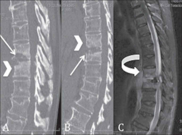
Low back pain is often managed at all levels of healthcare. In general, diagnostic investigation begins with radiography of the lumbar spine. In addition to the most common findings, radiologists can identify increased density of a vertebral body, referred to as ivory vertebra. The objective of this study was to describe the main diseases that can present with this radiologic sign, such as Hodgkin lymphoma, Paget's disease, metastatic prostate cancer, breast cancer, and osteomyelitis. It is extremely important that radiologists be aware of this finding in order to inform the requesting physician of the possible etiologies, given that it can be the initial radiologic presentation for these diseases.
Pacientes com dor lombar são frequentemente atendidos em todos os níveis de saúde. O início da investigação diagnóstica, de modo geral, se dá com a realização das radiografias da coluna lombar. Além dos achados mais frequentes, os profissionais podem encontrar uma vértebra de densidade muito aumentada em comparação com as demais, chamada de vértebra em marfim. O objetivo deste trabalho é descrever as principais doenças que podem apresentar, entre suas características radiológicas, este tipo de vértebra, como linfoma de Hodgkin, doença de Paget, metástases de neoplasias prostáticas e de mama, além da osteomielite. Por ser, em alguns casos, a apresentação radiológica inicial dessas doenças, é de suma importância que o radiologista tenha conhecimento deste achado e oriente o profissional solicitante quanto às suas possíveis causas.
Keywords: Lymphoma; Neoplasm metastasis; Osteitis deformans; Osteomyelitis; Spine.
Figures
References
-
- Terazaki CRT, Trippia CR, Trippia CH, et al. Synovial chondromatosis of the shoulder: imaging findings. Radiol Bras. 2014;47:38–42.
-
- Arend CF. Sonography of the iliotibial band: spectrum of findings. Radiol Bras. 2014;47:33–37.
-
- Nakamura SA, Lorenzato MM, Engel EE, et al. Incidental enchondromas at knee magnetic resonance imaging: intraobserver and interobserver agreement and prevalence of imaging findings. Radiol Bras. 2013;46:129–133.
LinkOut - more resources
Full Text Sources
Other Literature Sources