

doi: 10.2484/rcr.v9i1.908.
eCollection 2014.
CNS toxoplasmosis in an immunocompetent individual
Affiliations
- PMID: 27141248
- PMCID: PMC4838758
- DOI: 10.2484/rcr.v9i1.908
Item in Clipboard
CNS toxoplasmosis in an immunocompetent individual
Radiol Case Rep.
.
Abstract
Toxoplasmosis is a serious and life-threatening disease in humans with a high prevalence in immunocompromised persons. The disease has a wide spectrum, depending on the immune status of the person. A CNS manifestation of toxoplasmosis in an immunocompetent person is very rare and often undetected. Our case of CNS toxoplasmosis in an immunocompetent person emphasizes the radiological diagnosis, which was further confirmed by advanced microbiology technique.
Figures
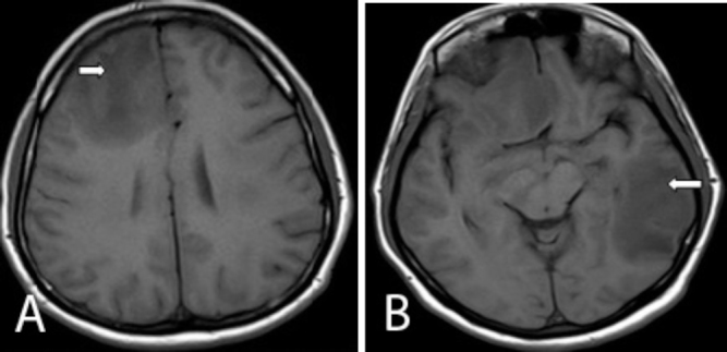
Axial T1 images of the brain show multiple hypointense lesions involving the right frontal (right arrow) and left temperoparietal lobes (left arrow).
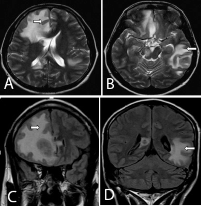
Axial T2 and coronal FLAIR images of the brain show multiple hyperintense lesions involving the right frontal (right arrow) and the left temperoparietal lobes (left arrow).
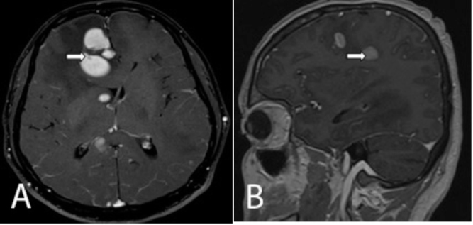
Axial and sagittal postcontrast T1W images show intensely enhancing lesions involving the bilateral cerebral hemispheres (right arrow),
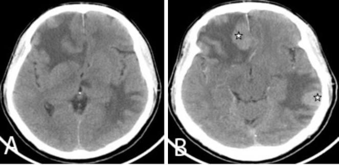
Axial pre- and postcontrast CT brain images show multiple intensely enhancing lesions (star) involving the bilateral cerebral hemispheres.
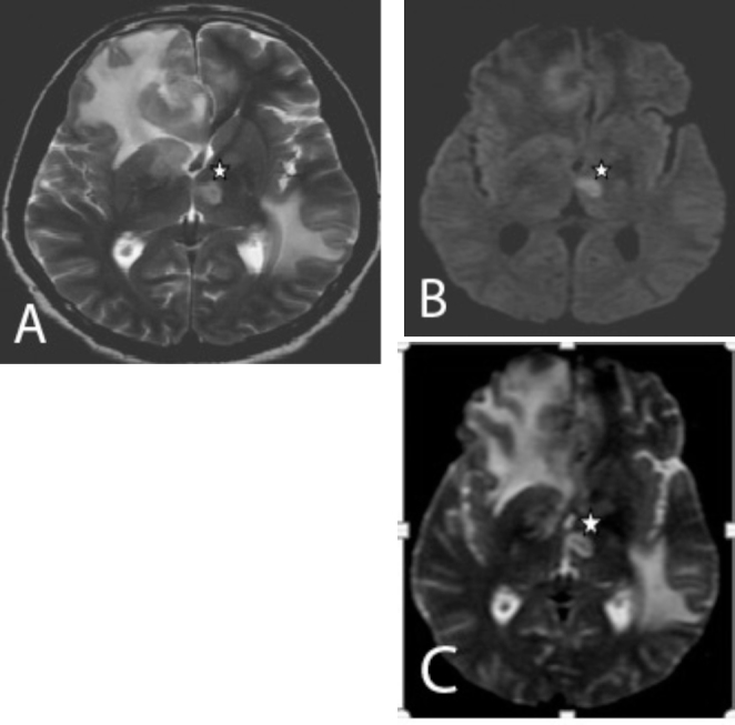
A and B. Axial T2W and DWI images show new hyperintense lesions with restricted diffusion in the left thalamus and the globus pallidus (star). C. Axial ADC image shows low signal in the left thalamus, suggestive of true restriction.
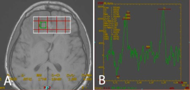
MR spectroscopy of the lesion involving the right frontal lobe shows elevated lipid lactate peak.
References
-
- Kornienko VN, Pronin IN. Diagnostic neuroradiology. Springer Verlag; 2009.
-
- Venkatasatya SA, Kumar GA, Pratap VS, Vivekananda DV, Madhukar R, Shyam S. Neurological manifestations in HIV infected patients around Varnasi, India. African Journal of Neuroscience. 2006;25:33–40.
-
- Carme B., Bissuel F., Ajzenberg D., Bouyne R., Aznar C., Demar M., Bichat S., Louvel D., Bourbigot A.M., Peneau C., Neron P., Dardé M.L. Severe acquired toxoplasmosis in immunocompetent adult patients in French Guiana. Journal of clinical microbiology. Nov. 2002;40:4037–4044. [PubMed] No. 11 0095-1137/02/$04.000. - PMC - PubMed
LinkOut - more resources
Full Text Sources
Other Literature Sources
