. 2016 Apr 14:7:66.
doi: 10.4103/2008-7802.180412. eCollection 2016.

Effect of Thymoquinone on P53 Gene Expression and Consequence Apoptosis in Breast Cancer Cell Line

Affiliations

Effect of Thymoquinone on P53 Gene Expression and Consequence Apoptosis in Breast Cancer Cell Line

Mehdi Nikbakht Dastjerdi et al. Int J Prev Med. .

Abstract

Background: Nigella sativa has been a nutritional flavoring factor and natural treatment for many ailments for so many years in medical science. Earlier studies have been reported that thymoquinone (TQ), an active compound of its seed, contains anticancer properties. Previous studies have shown that TQ induces apoptosis in breast cancer cells but it is unclear the role of P53 in the apoptotic pathway. Hereby, this study reports the potency of TQ on expression of tumor suppressor gene P53 and apoptosis induction in breast cancer cell line Michigan Cancer Foundation-7 (MCF-7).

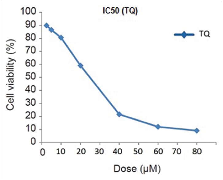
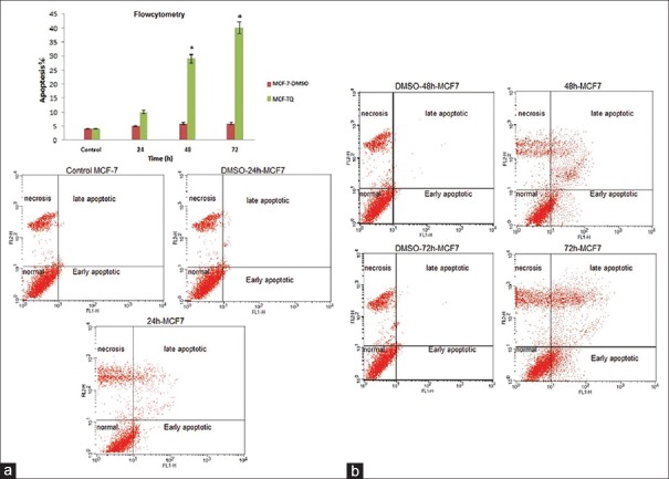
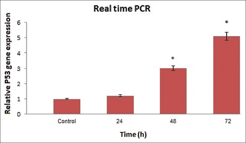
Methods: MCF-7 cell line was cultured and treated with TQ, and 3-(4,5-dimethylthiazol-2-yl)-2,5-diphenyltetrazolium bromide (MTT) assay was carried out for evaluating the half-maximal inhibitory concentration (IC50) values after 24 h of treatment. The percentage of apoptotic cells was measured by flow cytometry. Real-time polymerase chain reaction (PCR) was performed to estimate the messenger RNA expression of P53 in MCF-7 cell line at different times.

Results: The IC50 value for the TQ in MCF-7 cells was 25 μM that determined using MTT assay. The flow cytometry and real-time PCR results showed that TQ could induce apoptosis in MCF-7 cells, and the P53 gene expression was dramatically up-regulated by ascending time, respectively. Hence, there was significant difference in 48 and 72 h.

Conclusions: Our results demonstrated that TQ could induce apoptosis in MCF-7 cells through up-regulation of P53 expression in breast cancer cell line (MCF-7) by time-dependent manner.

Keywords: Apoptosis; Michigan C]ancer Foundation-7 cells; genes P53; thymoquinone.


Figures

Figure 1
Figure 1
Half-maximal inhibitory concentration assay for half-maximal inhibitory concentration analysis of thymoquinone in Michigan Cancer Foundation-7 cancer cell lines after 24 h of treatment. Cells were incubated with or without the thymoquinone using 25 μM dose. The relative amount of viable cells was estimated by measuring the absorbance of the cell suspension after incubation with 3-(4,5-dimethylthiazol-2-yl)-2,5-diphenyltetrazolium bromide assay. A graph of viability versus drug concentration was used to calculate half-maximal inhibitory concentration values for Michigan Cancer Foundation-7 cell line
Figure 2
Figure 2
(a) Relative levels of apoptotic cells in Michigan Cancer Foundation-7 treated with thymoquinone for different times. Cells incubated with the vehicle dimethyl sulfoxide were used as a control. The percentage of apoptotic cells was measured using the Annexin V-FITC flow cytometry 1-histogram and propidium iodide flow cytometry 2-histogram. Dunnett's test was used for considered comparisons (*P < 0.05) (in any figure, right upper quarter show late apoptotic cells, left upper quarter show necrosis cells, right lower quarter show early apoptotic cells and left lower quarter show normal cells). (b) Cells that are Annexin V-positive and propidium iodide negative are in early apoptosis as phosphatidyl serine translocation has occurred; although, the plasma membrane remains intact. Cells that are positive for both Annexin V and propidium iodide either are in the late stages of apoptosis or are already dead as phosphatidyl serine translocation has occurred and the loss of plasma membrane integrity is visible. *P < 0.05 compared to controls. Dunnett's test was used for considered comparisons (*P < 0.05)
Figure 3
Figure 3
Results of real-time quantitative polymerase chain reaction before and after thymoquinone treatment at different times on the P53 messenger RNA expression in Michigan Cancer Foundation-7 cell. Relative expression levels were obtained using the comparative Ct (ΔΔCt) method (*P < 0.05)

Similar articles

Cited by

References

    1. Autier P, Boniol M, Gavin A, Vatten LJ. Breast cancer mortality in neighbouring European countries with different levels of screening but similar access to treatment: Trend analysis of WHO mortality database. BMJ. 2011;343:d4411. - PMC - PubMed
    1. Montazeri A, Vahdaninia M, Harirchi I, Harirchi AM, Sajadian A, Khaleghi F, et al. Breast cancer in Iran: Need for greater women awareness of warning signs and effective screening methods. Asia Pac Fam Med. 2008;7:6. - PMC - PubMed
    1. Coley HM. Mechanisms and strategies to overcome chemotherapy resistance in metastatic breast cancer. Cancer Treat Rev. 2008;34:378–90. - PubMed
    1. Burits M, Bucar F. Antioxidant activity of Nigella sativa essential oil. Phytother Res. 2000;14:323–8. - PubMed
    1. Ravindran J, Nair HB, Sung B, Prasad S, Tekmal RR, Aggarwal BB. Thymoquinone poly (lactide-co-glycolide) nanoparticles exhibit enhanced anti-proliferative, anti-inflammatory, and chemosensitization potential. Biochem Pharmacol. 2010;79:1640–7. - PMC - PubMed