. 2015 Oct 29;5(3):e1091555.
doi: 10.1080/2162402X.2015.1091555. eCollection 2016 Mar.

Characterization of the first-in-class T-cell-engaging bispecific single-chain antibody for targeted immunotherapy of solid tumors expressing the oncofetal protein claudin 6

Affiliations

Characterization of the first-in-class T-cell-engaging bispecific single-chain antibody for targeted immunotherapy of solid tumors expressing the oncofetal protein claudin 6

Christiane R Stadler et al. Oncoimmunology. .

Abstract

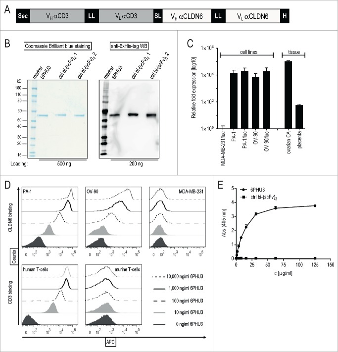
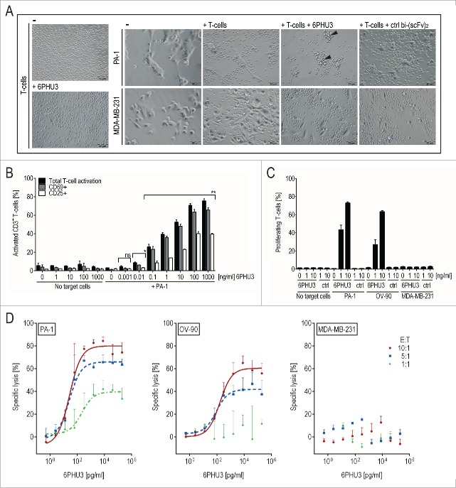
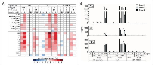
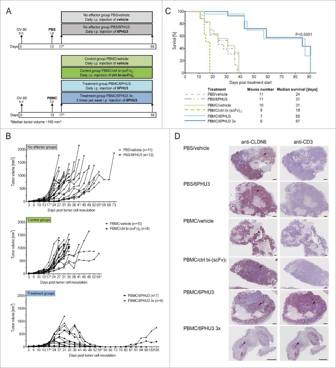
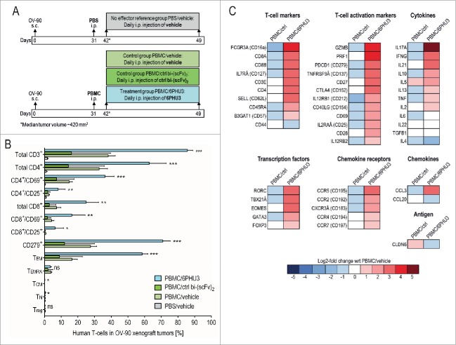
The fetal tight junction molecule claudin 6 (CLDN6) is virtually absent from any normal tissue, whereas it is aberrantly and frequently expressed in various cancers of high medical need. We engineered 6PHU3, a T-cell-engaging bispecific single chain molecule (bi-(scFv)2) with anti-CD3/anti-CLDN6 specificities, and characterized its pharmacodynamic properties. Our data show that upon engagement by 6PHU3, T cells strongly upregulate cytotoxicity and activation markers, proliferate and acquire an effector phenotype. 6PHU3 exerts potent killing of cancer cells in vitro with EC50 values in the pg/mL range. Subcutaneous xenograft tumors in NSG mice engrafted with human PBMCs are eradicated by 6PHU3 treatment and survival of mice is significantly prolonged. Tumors of 6PHU3-treated mice are strongly infiltrated with activated CD4+, CD8+ T cells and TEM type cells but not Tregs and display a general activation of a mostly inflammatory phenotype. These effects are only observed upon bispecific but not monospecific engagement of 6PHU3. Together with the exceptionally cancer cell selective expression of the oncofetal tumor marker CLDN6, this provides a safeguard with regard to toxicity. In summary, our data shows that the concept of T-cell redirection combined with that of highly selective targeting of CLDN6-positive solid tumors is effective. Thus, exploring 6PHU3 for clinical therapy is warranted.

Keywords: Bispecific antibody; T cell engagement; T-cell engager; ideal target; oncofetal tumor marker; solid tumors; targeted immunotherapy; tumor-infiltrating lymphocytes; xenograft mouse model.


Figures

Figure 1.
Figure 1.
6PHU3 binds selectively to CLDN6 and CD3. (A) Schematic overview of the bi-(scFv)2 6PHU3 sequence cassettes cloned into pcDNA3.1 mammalian expression vector. (B) SDS-PAGE analysis of IMAC-purified 6PHU3 and two different control bi-(scFv)2. (C) CLDN6 expression of human carcinoma cell lines PA-1(/luc), OV-90(/luc) and MDA-MB-231/luc as determined by qPCR. Fold expression of CLDN6 expression has been calculated from two independent experiments. Tissue samples from ovarian carcinoma and placenta served as positive controls, CLDN6 tissues heart and thymus as calibrators. (D) 6PHU3 target binding as measured by flow cytometry. Increasing concentrations of 6PHU3 were incubated with CLDN6+ cells (PA-1, OV-90) or CD3+ human T-cells. MDA-MB-231/luc cells and murine T cells served as negative controls. Bound proteins were detected via their 6xHis-tag. (E) Bispecificity of 6PHU3 as demonstrated by ELISA. 6PHU3 and a control bi-(scFv)2 were captured by a CD3-mimicking peptide. Detection was conducted by an antibody specific for the anti-CLDN6 scFv, and a 2nd detection antibody. ABS indicates absorption; APC, allophycocyanin; bi-(scFv)2, bispecific single chain variable fragment; CA, carcinoma; c, concentration; ctrl, control; H, 6xHis-tag; hu, human; LL, long linker (15–18 amino acids); mu, murine; Sec, secretion signal; SL, short linker (five amino acids); VL, variable light chain; VH, variable heavy chain; WB, western blot.
Figure 2.
Figure 2.
6PHU3 mediates strong T-cell-engaging effects only in the presence of target cells. (A) Microscopic image after 24 h of co-culturing CLDN6+ PA-1/luc cells and CLDN6 MDA-MB-231/luc control cells in an E:T ratio of 5:1 +/− 30 ng/mL 6PHU3 or ctrl bi-(scFv)2 as indicated (200× magnification). Arrowheads: clusters of T cells on residual target cells (B) Activation of CD3+ T cells as analyzed by flow cytometry. T cells were incubated with the indicated 6PHU3 concentrations +/− PA-1 cells for 48 h. Percentages of total activated T cells (black columns), CD69+ (gray columns) and CD25+ (white columns) are shown. Ns signifies not significant (p = 0.053); *p < 0.002; **p < 0.0001. (C) Proliferation of CD45+ T-cells as determined by flow cytometry. CFSE-labeled T cells were seeded alone, with PA-1, OV-90 or MDA-MB-231/luc control cells in an E:T ratio of 10:1. Cells were stimulated with bi-(scFv)2 as indicated for 72 h. (D) Specific lysis of 6PHU3 as determined by a luciferase detection cytotoxicity assay. PBMC of three independent donors were co-incubated with CLDN6+ target cells PA-1/luc (left) or OV-90/luc (middle) and with CLDN6 cells MDA-MB-231/luc (right) in E:T ratios of 10:1 (red solid curves), 5:1 (blue dashed curves) and 1:1 (green dotted curve) for 48 h. Curves present the average specific lysis by three donors, error bars show the deviation between donors. A 9-fold serial dilution of 6PHU3 (0.0005–200 ng/mL) was applied.
Figure 3.
Figure 3.
T cells activated by 6PHU3 present a cytotoxic, pro-inflammatory phenotype. T cells were incubated with 5 or 500 ng/mL 6PHU3 +/− PA-1 cells or MDA-MB-231/luc control cells. (A) Heatmap showing log2-fold expression with respect to untreated T cells. Gene expression was evaluated using a custom-made panel of commercial primers specific for human T-cell identification-markers as measured by Fluidigm. All values were normalized to CD3. (B) T-cell cytokine release as measured by ELISA. Wrt indicates with respect to.
Figure 4.
Figure 4.
6PHU3 treatment eradicates tumors and extends survival in a xenograft mouse model. (A) Injection schedule scheme. Mice with advanced OV-90 xenografts were subjected to the following treatments: ‘No effector’ groups were not engrafted with human PBMC but treated daily with either vehicle or 6PHU3. ‘Control’ groups were engrafted with PBMC and treated daily with either vehicle or the control bi-(scFv)2. ‘Treatment’ groups underwent PBMC grafting and were treated with 6PHU3 according to two different schedules. (B) Tumor growth of all mice and groups. Treatment was applied i.p. between the days marked by an asterisk (*). Each line represents an individual mouse. (C) Kaplan–Meier survival curves of all groups from the day of treatment start. (D) Immunohistochemistry of representative xenograft whole tumor sections. Black arrow heads point to example areas of reddish-brown staining indicating the presence of CLDN6 (left) or CD3 (right) in adjacent sections. Black dashes correlate to 1 mm of tumor size. I.p. indicates intraperitoneal; s.c., subcutaneous.
Figure 5.
Figure 5.
T cells infiltrated into subcutaneous xenograft tumors in response to 6PHU3 have an activated and cytotoxic functional status. (A) Injection schedule scheme. (B) Flow cytometric analysis of human T cells in whole tumor tissue. Percentage of TILs from gated live singlet total cells is depicted. Significance was calculated by comparing PBMC/6PHU3 to PBMC/ctrl bi-(scFv)2 and to PBMC/vehicle. (C) Heatmap showing a log2-fold expression of PBMC/ctrl bi-(scFv)2 (‘PBMC/ctrl’) and PBMC/6PHU3 groups with respect to PBMC/vehicle group as reference (mean values of two independent runs). Gene expression was evaluated using a predesigned primer panel specific for 46 human T-cell identification-markers as measured by Fluidigm. Ns indicates not significant; TCM, central memory T cells; TEM, effector memory T cells; TEMRA, CD45RA+ effector memory T cells; TN, naive T cells; Treg, regulatory T cells; *p < 0.03; **p < 0.008; ***p < 0.0009.

Similar articles

Cited by

References

    1. Clark MR, Waldmann H. T-cell killing of target cells induced by hybrid antibodies: comparison of two bispecific monoclonal antibodies. J Natl Cancer Inst 1987; 79:1393-401; PMID:3121901 - PubMed
    1. Lanzavecchia A, Scheidegger D. The use of hybrid hybridomas to target human cytotoxic T lymphocytes. Eur J Immunol 1987; 17:105-11; PMID:3102250; http://dx.doi.org/10.1002/eji.1830170118 - DOI - PubMed
    1. Sahin U, Hartmann F, Senter P, Pohl C, Engert A, Diehl V, Pfreundschuh M. Specific activation of the prodrug mitomycin phosphate by a bispecific anti-CD30/anti-alkaline phosphatase monoclonal antibody. Cancer Res 1990; 50:6944-48; PMID:2170012 - PubMed
    1. Staerz UD, Kanagawa O, Bevan MJ. Hybrid antibodies can target sites for attack by T cells. Nature 1985; 314:628-31; PMID:2859527; http://dx.doi.org/10.1038/314628a0 - DOI - PubMed
    1. Ruf P, Gires O, Jäger M, Fellinger K, Atz J, Lindhofer H. Characterisation of the new EpCAM-specific antibody HO-3: implications for trifunctional antibody immunotherapy of cancer. Br J Cancer 2007; 97:315-21; PMID:17622246; http://dx.doi.org/10.1038/sj.bjc.6603881 - DOI - PMC - PubMed