. 2015 Dec 17;5(3):e1112941.
doi: 10.1080/2162402X.2015.1112941. eCollection 2016 Mar.

Antitumor cytotoxicity induced by bone-marrow-derived antigen-presenting cells is facilitated by the tumor suppressor protein p53 via regulation of IL-12

Affiliations

Antitumor cytotoxicity induced by bone-marrow-derived antigen-presenting cells is facilitated by the tumor suppressor protein p53 via regulation of IL-12

Tania L Slatter et al. Oncoimmunology. .

Abstract

Activated antigen-presenting cells (APC) deliver the three signals cytotoxic T cells require to differentiate into effector cells that destroy the tumor. These comprise antigen, co-stimulatory signals and cytokines. Once these cells have carried out their function, they apoptose. We hypothesized that the tumor suppressor protein, p53, played an important role in generating the antitumor response facilitated by APC. CD11c+ APC derived from p53 wild-type (wt) mouse (wt p53) GM-CSF bone marrow cultures (BMAPC) and activated had reduced survival compared to BMAPC from p53 null consistent with p53-mediated apoptosis following activation. There was a lower percentage of antigenic peptide/MHC I complexes on antigen-pulsed p53 null cells suggesting p53 played a role in antigen processing but there was no difference in antigen-specific T cell proliferative responses to these cells in vivo. In contrast, antigen-specific cytotoxicity in vivo was markedly reduced in response to p53 null BMAPC. When these cells were pulsed with a model tumor antigen and delivered as a prophylactic vaccination, they provided no protection against melanoma cell growth whereas wt BMAPC were very effective. This suggested that p53 might regulate the requisite third signal and, indeed, we found that p53 null BMAPC produced less IL-12 than wt p53 BMAPC and that p53 bound to the promoter region of IL-12. This work suggests that p53 in activated BMAPC is associated with the generation of IL-12 required for the differentiation of cytotoxic immune responses and an effective antitumor response. This is a completely new role for this protein that has implications for BMAPC-mediated immunotherapy.

Keywords: Cytotoxic immune response; dendritic cells; interleukin 12; p53 tumor suppressor protein; tumor vaccination.


Figures

Figure 1.
Figure 1.
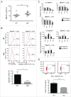
p53 is increased in activated wild-type BMAPC. (A) Activation markers are increased on wt p53 BMAPC after LPS treatment. Immature BMDC were activated with LPS (1 µg/mL) for 16 h or left unactivated (UA). Cells were stained with antibodies toward CD11c, and activation markers CD40 and CD86 or the corresponding isotype controls. The amount of CD40 and CD86 on CD11c positive cells was measured and compared with that on the isotype control stained cells using flow cytometry. Bar graph represents mean +/− SD n = 3. (B) p53 is increased in activated BMAPC. BMAPC were activated with LPS (+LPS; 1 µg/mL) overnight or left untreated (-LPS). Cells were stained with antibodies toward CD11c and p53. p53 was measured in CD11c positive cells using flow cytometry and the percentage of p53 positive cells compared between activated and untreated cells. An example of a flow cytometric histogram is given as well as quantitative data for percentage of p53 positive cells and mean fluorescence intensity (MFI) n = 6. (C) Western blot replicates of BMAPC showing p53 protein in LPS activated (+LPS) and unactivated BMAPC (-LPS). *p < 0.05; **p < 0.01; p < 0.001; ****p <0.0001 The ‘Control’, Lane 1, contains lysate from BMAPC treated with amsacrine, a topoisomerase II inhibitor.
Figure 2
Figure 2
BMAPC survival in vitro is p53 dependent, but the activation phenotype and antigen presentation is p53 independent. (A) The yield of BMAPC from bone marrow is reduced with wt p53. Bone marrow was isolated from wt p53 and p53 null mice and equal numbers of live cells (trypan blue negative) placed into cell culture to prepare BMAPC. At day 6, the percentage of CD11c positive cells was counted using flow cytometry and compared between p53 genotypes. (B) Apoptotic cell death is increased in wt p53 BMAPC. BMAPC from wt p53 and p53 null mice were treated overnight with CpG (1 µg/mL), LPS (1 µg/mL) or left untreated. The apoptotic cell detection reagent FLICA was added 30 min before cells were harvested. Harvested cells were stained with CD11c and the amount of apoptotic cells (FLICA positive) in the CD11c positive population measured using flow cytometry and compared between p53 genotypes (p = 0.0068, n = 3 per genotype). (C) p53 null BMAPC respond to activation signals by upregulating cell surface co-stimulatory molecules similar to wt p53 BMAPC. BMAPC from wt p53 and p53 null mice were activated overnight with LPS (5 µg/mL), CpG (1 µg/mL) or left untreated (UT). Cells were harvested and stained with antibodies toward CD11c, and co-stimulatory markers MHC11, CD80, CD86 and CD40 or the corresponding isotype control. Each marker was analyzed in CD11c positive cells using flow cytometry. The percentage of positive cells (top panel) or the mean fluorescence intensity (bottom panel) for each marker was compared between genotypes with no statistical differences found (results, mean ± SD, n = 3 per genotype). (D) p53 null BMAPC presented less ovalbumin peptide complexed to MHCI (MHCI/OVA peptide) than wt p53 cells. BMAPC from wt p53 and p53 null mice were pulsed with ovalbumin conjugated to virus like particles (VLP OVA). Cells were harvested, stained, and gated on CD11c positive cells and then stained with antibody to detect MHCI/SIINFEKL peptide complexes (or an isotype control). CD11c cells positive for MHCI/SIINFEKL peptide complexes were detected using flow cytometry (E) Mean percentage MHCI/OVA peptide +/− SD n3 (F). *; ***p < 0.001.
Figure 3.
Figure 3.
Survival of BMAPC in vivo is p53 dependent. (A) wt p53 adoptively transferred BMAPC have a greater percentage of apoptotic cells compared with p53 null BMAPC. BMAPC from wt p53 and p53 null mice were injected intra-dermally into wt p53 hosts. Skin was harvested 4 h post injection in close proximity to the injection site (within 0.2 mm) and stained with an antibody to detect active caspase 3 to identify apoptotic cells. Positive cells were identified using DAB and light microcopy (top panel) and the percentage of positive cells per 200 total cells was calculated and compared between genotypes (bottom panel: results, mean ± SD, n= 3 per genotype). (B) p53 null and wt p53 BMAPC express the receptor for pro-migratory chemokines, CCR7. BMDC from wt p53 and p53 null mice were activated with CpG (1 µg/mL) or left untreated. Cells were harvested and stained with antibodies toward CD11c and CCR7. The percentage of CD11c cells positive for CCR7 in activated cells is compared with that in untreated (UA), and cells stained with the isotype control using flow cytometry (Top panel). Mean fluorescence intensity of CCR7 expression is compared between p53 genotypes with and without CpG activation (bottom panel, results mean ± SD, n=3 per genotype; *p < 0.05). (C) The migration of BMAPC to draining lymph nodes is increased in p53 null BMAPC. BMAPC from wt p53 and p53 null mice were labeled with different concentrations of CFSE (wt p53 BMDC were labeled with a higher CFSE concentration compared to p53 null BMDC). Draining lymph nodes were harvested 8–72 h after injection and the percentage of CFSE positive cells measured by flow cytometry and compared between genotypes. Results, mean ± sd, n = 3 per genotype; *p < 0.05; **p < 0.01; ***p < 0.001.
Figure 4.
Figure 4.
T cell proliferation is p53 independent, however, for an effective cytotoxic T cell response p53 is required. (A) p53 null BMAPC-induced T cell proliferation similar to wt p53 BMAPC. OTI CD8+ T cells labeled with CFSE were adoptively transferred to wt p53 recipient mice. One day later, BMAPC from wt p53 and p53 null mice pulsed with SIINFEKL peptide were injected intra-dermally into the recipient mice on opposite flanks. 72 h later, draining lymph nodes were removed and CFSE labeled cells measured using flow cytometry and compared between genotypes (the results from eight recipient mice (1–8) are shown). (B) wt p53 BMAPC induces a cytotoxic T cell response more effectively. wt p53 recipient mice were vaccinated with BMAPC from wt p53 or p53 null mice pulsed with ovalbumin (OVA), SIINFEKL peptide, or were left untreated. One-week later, mice were injected with wt p53 splenocytes pulsed with SIINFEKL peptide and labeled with a high concentration of CFSE at a 1:1 ratio with splenocytes not pulsed with antigen and labeled with a low concentration of CFSE. Blood and spleen of recipient mice were collected and CFSE high and low dose labeled cells identified by flow cytometry. The percentage of specific lysis was calculated (material and methods) and compared between genotypes (results, mean ±SEM, n = 3 per genotype). *p < 0.05; **p < 0.01; ****p < 0.0001.
Figure 5.
Figure 5.
Protection against tumor development by BMAPC vaccination depends on p53 function. Mice were prophylactically injected with BMAPC from wt p53 or p53 null mice pulsed with antigen (ovalbumin (OVA), or SIINFEKL), without antigen (UT), or the PBS vehicle alone (B16OVA). One week later, these mice were implanted with B.16OVA melanoma cells subcutaneously. Mice were monitored daily for tumor development and sacrificed when the tumor reached 15 mm2 in diameter. Survival times between animal groups were compared (n = 10 per group). **comparison between wt p53 and p53 null BMAPC pulsed with OVA; *comparison between wt p53 and p53 null BMAPC pulsed with SIINFEKL.
Figure 6.
Figure 6.
IL-12 production is higher and transcription occurs earlier in wt p53 activated BMAPC. (A, B) IL-12p40/70 is increased in wt p53 BMAPC following CpG activation. BMAPC from wt p53 and p53 null mice were activated with CpG (1 µg/mL) or left untreated. Cells were harvested and stained with antibodies toward CD11c and IL12p40/70. Positive cells were identified by flow cytometry (upper panel) and the percentage of IL-12p40/70 positive cells compared between genotypes (lower panel: results, mean ± SD n = 3 per genotype). *p < 0.05; ***p < 0.001. (C) IL-12 is increased in BMAPC supernatants from wt p53 mice. wt p53 and p53 null BMDC were activated with LPS (1 µg/mL) or left untreated (UT), cultured for 24–120 h, and IL-12 measured in supernatants by ELISA (results, mean ± SD n=4 per genotype) *p <0.05; **p < 0.01. (D) Il12b is expressed earlier in wt p53 BMAPC following CpG activation. BMAPC from wt p53 and p53 null mice were activated with CpG (1 µg/mL) or left untreated. Cells were harvested 30 min to 2 h post activation and the quantity of the Il12b transcript measured by real time PCR and expressed as the fold difference compared with three housekeeping genes measured. (E) p53 binds to the IL-12p40 promoter. Chromatin immunopreciptation assay on wt p53 (+/+IP) and null MEFS with (−/−IP) pre-immune cleared with normal rabbit serum and Protein G beads (negative); (+/+ Input; −/− Input = 5% of lysate subjected to IP).

Similar articles

Cited by

References

    1. Kushwah R, Hu J. Dendritic cell apoptosis: regulation of tolerance vs. immunity. J Immunol 2010; 185:795-802; PMID:20601611; http://dx.doi.org/2063680510.4049/jimmunol.1000325. - DOI - PubMed
    1. Chen M, Wang J. Programmed cell death of dendritic cells in immune regulation. Immunol Rev 2010; 236:11-27; PMID:20636805; http://dx.doi.org/10.1111/j.1600-065X.2010.00916.x. - DOI - PMC - PubMed
    1. Oren M. Decision making by p53: life, death and cancer. Cell Death Diff 2003; 10:431-42; PMID:12719720; http://dx.doi.org/10.1038/sj.cdd.4401183. - DOI - PubMed
    1. Braithwaite AW, Royds JA, Jackson P. The p53 story: layers of complexity. Carcinogenesis 2005; 26:1161-9; PMID:15817608; http://dx.doi.org/10.1093/carcin/bgi091. - DOI - PubMed
    1. Lowe SW, Jacks T, Housman DE, Ruley HE. Abrogation of oncogene-associated apoptosis allows transformation of p53-deficient cells. Proc Natl Acad Sci U S A 1994; 91:2026-30; PMID:8134344; http://dx.doi.org/10.1073/pnas.91.6.2026. - DOI - PMC - PubMed

Publication types