

Oncolytic adenoviruses coated with MHC-I tumor epitopes increase the antitumor immunity and efficacy against melanoma
- PMID: 27141389
- PMCID: PMC4839367
- DOI: 10.1080/2162402X.2015.1105429
Oncolytic adenoviruses coated with MHC-I tumor epitopes increase the antitumor immunity and efficacy against melanoma
Abstract
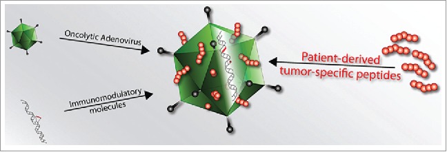
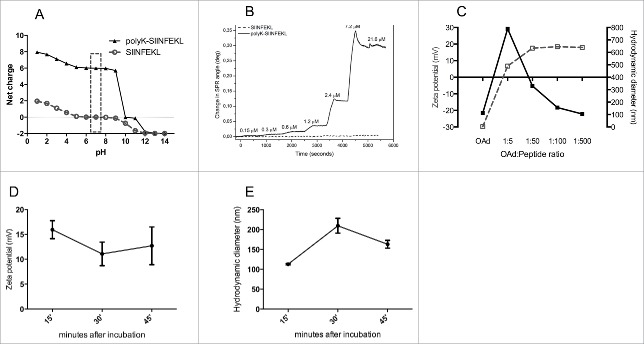
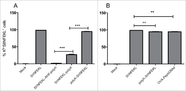
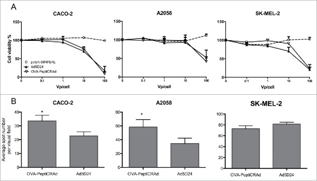
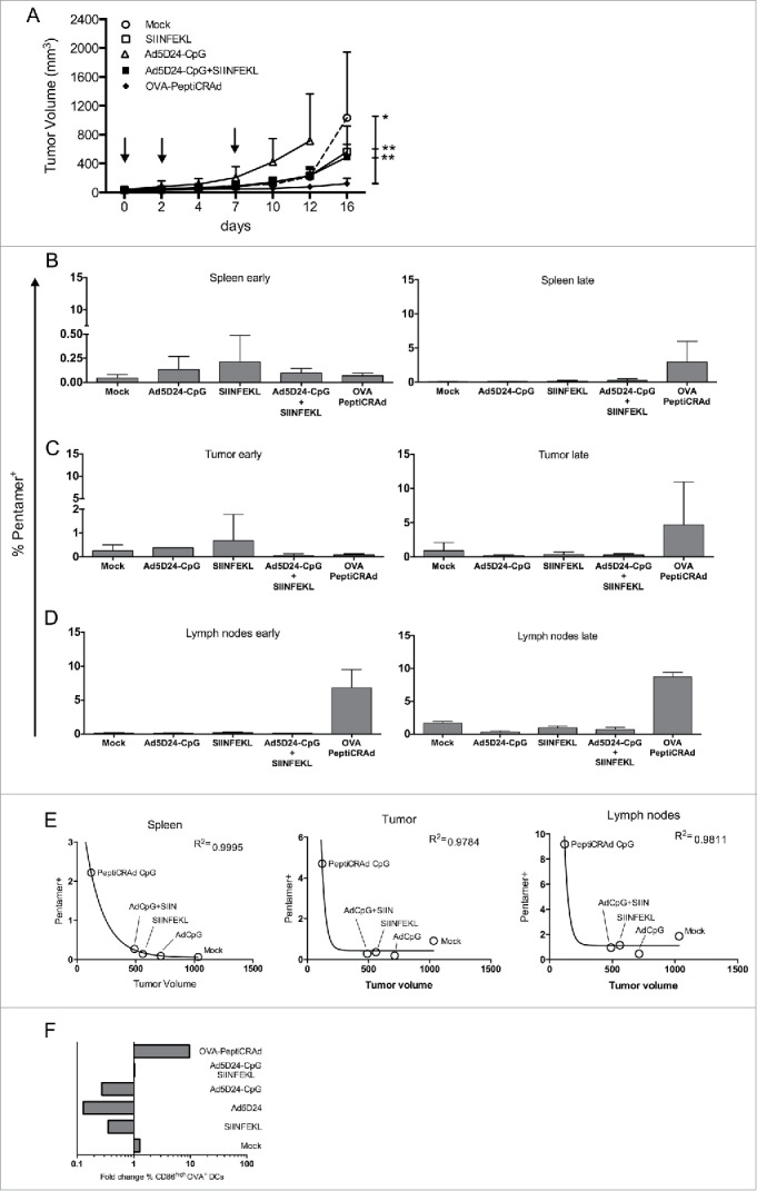
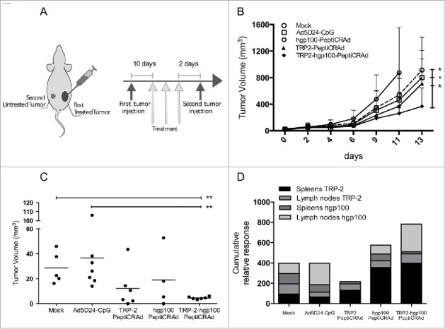
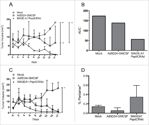
The stimulation of the immune system using oncolytic adenoviruses (OAds) has attracted significant interest and several studies suggested that OAds immunogenicity might be important for their efficacy. Therefore, we developed a versatile and rapid system to adsorb tumor-specific major histocompatibility complex class I (MHC-I) peptides onto the viral surface to drive the immune response toward the tumor epitopes. By studying the model epitope SIINFEKL, we demonstrated that the peptide-coated OAd (PeptiCRAd) retains its infectivity and the cross presentation of the modified-exogenous epitope on MHC-I is not hindered. We then showed that the SIINFEKL-targeting PeptiCRAd achieves a superior antitumor efficacy and increases the percentage of antitumor CD8+ T cells and mature epitope-specific dendritic cells in vivo. PeptiCRAds loaded with clinically relevant tumor epitopes derived from tyrosinase-related protein 2 (TRP-2) and human gp100 could reduce the growth of primary-treated tumors and secondary-untreated melanomas, promoting the expansion of antigen-specific T-cell populations. Finally, we tested PeptiCRAd in humanized mice bearing human melanomas. In this model, a PeptiCRAd targeting the human melanoma-associated antigen A1 (MAGE-A1) and expressing granulocyte and macrophage colony-stimulating factor (GM-CSF) was able to eradicate established tumors and increased the human MAGE-A1-specific CD8+ T cell population. Herein, we show that the immunogenicity of OAds plays a key role in their efficacy and it can be exploited to direct the immune response system toward exogenous tumor epitopes. This versatile and rapid system overcomes the immunodominance of the virus and elicits a tumor-specific immune response, making PeptiCRAd a promising approach for clinical testing.
Keywords: Cancer vaccine; humanized mice; immunotherapy; melanoma; oncolytic adenovirus; oncolytic vaccine; tumor epitopes.
Figures
Similar articles
-
Detection of naturally processed and HLA-A1-presented melanoma T-cell epitopes defined by CD8(+) T-cells' release of granulocyte-macrophage colony-stimulating factor but not by cytolysis.Clin Cancer Res. 1996 Jan;2(1):87-95. Clin Cancer Res. 1996. PMID: 9816095
-
Exploiting Preexisting Immunity to Enhance Oncolytic Cancer Immunotherapy.Cancer Res. 2020 Jun 15;80(12):2575-2585. doi: 10.1158/0008-5472.CAN-19-2062. Epub 2020 Feb 27. Cancer Res. 2020. PMID: 32107211
-
Peptides-Coated Oncolytic Vaccines for Cancer Personalized Medicine.Front Immunol. 2022 Apr 14;13:826164. doi: 10.3389/fimmu.2022.826164. eCollection 2022. Front Immunol. 2022. PMID: 35493448 Free PMC article.
-
Oncolytic Adenovirus: Prospects for Cancer Immunotherapy.Front Microbiol. 2021 Jul 21;12:707290. doi: 10.3389/fmicb.2021.707290. eCollection 2021. Front Microbiol. 2021. PMID: 34367111 Free PMC article. Review.
-
Showing the Way: Oncolytic Adenoviruses as Chaperones of Immunostimulatory Adjuncts.Biomedicines. 2016 Sep 19;4(3):23. doi: 10.3390/biomedicines4030023. Biomedicines. 2016. PMID: 28536390 Free PMC article. Review.
Cited by
-
Exploring the DNA2-PNA heterotriplex formation in targeting the Bcl-2 gene promoter: A structural insight by physico-chemical and microsecond-scale MD investigation.Heliyon. 2024 Jan 22;10(3):e24599. doi: 10.1016/j.heliyon.2024.e24599. eCollection 2024 Feb 15. Heliyon. 2024. PMID: 38317891 Free PMC article.
-
Low-dose decitabine enhances the efficacy of viral cancer vaccines for immunotherapy.Mol Ther Oncol. 2024 Jan 26;32(1):200766. doi: 10.1016/j.omton.2024.200766. eCollection 2024 Mar 21. Mol Ther Oncol. 2024. PMID: 38596301 Free PMC article.
-
The Benefit of Reactivating p53 under MAPK Inhibition on the Efficacy of Radiotherapy in Melanoma.Cancers (Basel). 2019 Aug 1;11(8):1093. doi: 10.3390/cancers11081093. Cancers (Basel). 2019. PMID: 31374895 Free PMC article.
-
A novel immunopeptidomic-based pipeline for the generation of personalized oncolytic cancer vaccines.Elife. 2022 Mar 22;11:e71156. doi: 10.7554/eLife.71156. Elife. 2022. PMID: 35314027 Free PMC article.
-
Dose Effects of Recombinant Adenovirus Immunization in Rodents.Vaccines (Basel). 2019 Oct 10;7(4):144. doi: 10.3390/vaccines7040144. Vaccines (Basel). 2019. PMID: 31658786 Free PMC article.
References
-
- Bischoff JR, Kirn DH, Williams A, Heise C, Horn S, Muna M, Ng L, Nye JA, Sampson-Johannes A, Fattaey A et al.. An adenovirus mutant that replicates selectively in p53-deficient human tumor cells. Science 1996; 274:373-6; PMID:8832876; http://dx.doi.org/10.1126/science.274.5286.373 - DOI - PubMed
-
- Heise C, Hermiston T, Johnson L, Brooks G, Sampson-Johannes A, Williams A, Hawkins L, Kirn D. An adenovirus E1A mutant that demonstrates potent and selective systemic anti-tumoral efficacy. Nat Med 2000; 6:1134-9; PMID:11017145; http://dx.doi.org/10.1038/80474 - DOI - PubMed
-
- Zhang SN, Choi IK, Huang JH, Yoo JY, Choi KJ, Yun CO. Optimizing DC vaccination by combination with oncolytic adenovirus coexpressing IL-12 and GM-CSF. Mol Ther 2011; 19:1558-68; PMID:21468000; http://dx.doi.org/10.1038/mt.2011.29 - DOI - PMC - PubMed
-
- Seiler MP, Gottschalk S, Cerullo V, Ratnayake M, Mane VP, Clarke C, Palmer DJ, Ng P, Rooney CM, Lee B. Dendritic cell function after gene transfer with adenovirus-calcium phosphate co-precipitates. Mol Ther 2007; 15:386-92; PMID:17235318; http://dx.doi.org/10.1038/sj.mt.6300029 - DOI - PubMed
-
- Cerullo V, Pesonen S, Diaconu I, Escutenaire S, Arstila PT, Ugolini M, Nokisalmi P, Raki M, Laasonen L, Sarkioja M et al.. Oncolytic adenovirus coding for granulocyte macrophage colony-stimulating factor induces antitumoral immunity in cancer patients. Cancer Res 2010; 70:4297-309; PMID:20484030; http://dx.doi.org/10.1158/0008-5472.CAN-09-3567 - DOI - PubMed
Publication types
Grants and funding
LinkOut - more resources
Full Text Sources
Other Literature Sources
Research Materials