. 2015 Dec 21;5(4):e1119354.
doi: 10.1080/2162402X.2015.1119354. eCollection 2016 Apr.

Dual targeting of glioblastoma with chimeric antigen receptor-engineered natural killer cells overcomes heterogeneity of target antigen expression and enhances antitumor activity and survival

Affiliations

Dual targeting of glioblastoma with chimeric antigen receptor-engineered natural killer cells overcomes heterogeneity of target antigen expression and enhances antitumor activity and survival

Sabrina Genßler et al. Oncoimmunology. .

Abstract

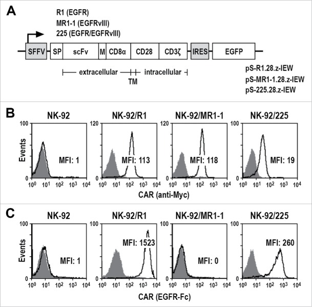
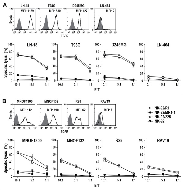
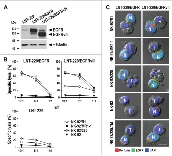
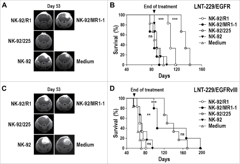
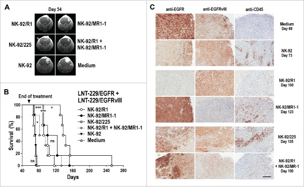
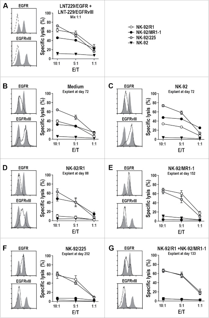
Epidermal growth factor receptor (EGFR) and its mutant form EGFRvIII are overexpressed in a large proportion of glioblastomas (GBM). Immunotherapy with an EGFRvIII-specific vaccine has shown efficacy against GBM in clinical studies. However, immune escape by antigen-loss variants and lack of control of EGFR wild-type positive clones limit the usefulness of this approach. Chimeric antigen receptor (CAR)-engineered natural killer (NK) cells may represent an alternative immunotherapeutic strategy. For targeting to GBM, we generated variants of the clinically applicable human NK cell line NK-92 that express CARs carrying a composite CD28-CD3ζ domain for signaling, and scFv antibody fragments for cell binding either recognizing EGFR, EGFRvIII, or an epitope common to both antigens. In vitro analysis revealed high and specific cytotoxicity of EGFR-targeted NK-92 against established and primary human GBM cells, which was dependent on EGFR expression and CAR signaling. EGFRvIII-targeted NK-92 only lysed EGFRvIII-positive GBM cells, while dual-specific NK cells expressing a cetuximab-based CAR were active against both types of tumor cells. In immunodeficient mice carrying intracranial GBM xenografts either expressing EGFR, EGFRvIII or both receptors, local treatment with dual-specific NK cells was superior to treatment with the corresponding monospecific CAR NK cells. This resulted in a marked extension of survival without inducing rapid immune escape as observed upon therapy with monospecific effectors. Our results demonstrate that dual targeting of CAR NK cells reduces the risk of immune escape and suggest that EGFR/EGFRvIII-targeted dual-specific CAR NK cells may have potential for adoptive immunotherapy of glioblastoma.

Keywords: Cetuximab; EGFR; EGFRvIII; chimeric antigen receptor; glioblastoma; natural killer cells.


Figures

Figure 1.
Figure 1.
Generation of CAR NK cells. (A) Lentiviral transfer plasmids pS-R1.28.z-IEW, pS-MR1-1.28.z-IEW and pS-225.28.z-IEW encoding under control of the Spleen Focus Forming Virus promoter (SFFV) CARs consisting of an immunoglobulin heavy chain signal peptide (SP), scFv fragments derived from EGFR-specific antibody R1, EGFRvIII-specific MR1-1, or 225 recognizing EGFR and EGFRvIII, followed by a Myc-tag (M), CD8α hinge region (CD8α), transmembrane and intracellular domains of CD28, and the intracellular domain of CD3ζ. Enhanced green fluorescent protein (EGFP) cDNA separated from the CAR sequence by an internal ribosome entry site (IRES) served as a marker. (B) CAR surface expression on NK-92/R1, NK-92/MR1-1 and NK-92/225 single cell clones was determined by flow cytometry with Myc-tag-specific antibody (open areas). Isotype antibody (filled areas) and parental NK-92 cells served as controls. (C) Binding of recombinant EGFR-Fc protein to the surface of CAR NK cells was measured by flow cytometry (open areas). CAR NK cells only treated with secondary antibody (filled areas) and parental NK-92 cells served as controls. MFI: mean fluorescence intensity (geometric mean).
Figure 2.
Figure 2.
Cytotoxicity of CAR NK cells against GBM cells. (A) Expression of EGFR on the surface of established LN-18, T98G, D245MG and LN-464 GBM cells was determined by flow cytometry with EGFR-specific antibody (open areas). Isotype antibody served as control (filled areas). Cell killing by NK-92/R1, NK-92/MR1-1 and NK-92/225 cells was investigated after co-incubation with the GBM cells for 2 h at different E/T ratios. Parental NK-92 were included for comparison. (B) Expression of EGFR on the surface of primary MNOF1300, MNOF132, R28 and RAV19 GBM cells and cell killing by CAR NK cells were determined as described above. Cytotoxicity data are shown as mean values ± SEM; n = 3. MFI: mean fluorescence intensity (geometric mean).
Figure 3.
Figure 3.
Cytotoxicity of CAR NK cells against EGFR- and EGFRvIII-expressing LNT-229 cells. (A) EGFR (170 kDa) and EGFRvIII (140 kDa) were detected in cell lysates of LNT-229 GBM cells ectopically overexpressing full-length EGFR (LNT-229/EGFR) or mutant EGFRvIII (LNT-229/EGFRvIII) by immunoblotting with an EGFR-specific antibody binding to both receptors. Parental LNT-229 served as control. (B) Cytotoxicity of CAR NK cells against LNT-229/EGFR, LNT-229/EGFRvIII and parental LNT-229 cells was investigated after co-incubation of effector and target cells for 2 h at different E/T ratios. Parental NK-92 were included as control. Mean values ± SEM are shown; n = 3. (C) Conjugate formation between CAR NK cells and LNT-229/EGFR and LNT-229/EGFRvIII cells was investigated by confocal microscopy. Tumor (T) and EGFP-positive CAR NK (N; green) cells were co-incubated for 1 h, fixed, permeabilized and stained for perforin (red) to identify cytotoxic granules. Cell nuclei were labeled with DAPI (blue). Parental NK-92 and NK-92/225.TM cells expressing a CAR without intracellular signaling domains were included as controls. Scale bar: 10 µm.
Figure 4.
Figure 4.
Antitumor activity of CAR NK cells against orthotopic LNT-229/EGFR and LNT-229/EGFRvIII GBM xenografts. (A) LNT-229/EGFR cells were stereotactically injected into the right striatum of NSG mice. Seven days later, the animals were treated by intratumoral injection of parental NK-92, EGFR-specific NK-92/R1, EGFRvIII-specific NK-92/MR1-1, or dual-specific NK-92/225 cells once per week for 12 weeks (n = 6). Control mice received injection medium. Tumor growth was monitored by MRI. Tumor development in representative animals from each group at day 53 is shown. (B) Symptom-free survival of the mice from the experiment described in (A). (C) LNT-229/EGFRvIII cells were stereotactically injected into the right striatum of NSG mice. Seven days later, the animals were treated as described above with NK-92 (n = 6), NK-92/R1 (n = 6), NK-92/MR1-1 (n = 5), or NK-92/225 (n = 6) cells once per week for 8 weeks. Control mice received injection medium (n = 6). Tumor development in representative animals from each group at day 53 is shown. (D) Symptom-free survival of the mice from the experiment described in (C). ***p ≤ 0.001; **p ≤ 0.01; ns, p > 0.05.
Figure 5.
Figure 5.
Antitumor activity of CAR NK cells against mixed LNT-229/EGFR and LNT-229/EGFRvIII GBM xenografts. (A) LNT-229/EGFR and LNT-229/EGFRvIII cells were mixed at a 1:1 ratio before stereotactic injection of the cells into the right striatum of NSG mice. Seven days later, the animals were treated by intratumoral injection of parental NK-92, EGFR-specific NK-92/R1, EGFRvIII-specific NK-92/MR1-1, dual-specific NK-92/225, or a 1:1 mixture of NK-92/R1 and NK-92/MR1-1 cells once per week for 8 weeks (n = 6). Control mice received injection medium. Tumor development in representative animals from each group at day 54 is shown. (B) Symptom-free survival of the mice from the experiment described in (A). ***p ≤ 0.001; *p ≤ 0.05; ns, p > 0.05. (C) Sections of tumors from individual animals of each treatment group sacrificed at the indicated time point were stained with EGFR- or EGFRvIII-specific antibodies. NK cells present in tumor tissues were detected with CD45-specific antibody. Scale bar: 300 µm.
Figure 6.
Figure 6.
Cytotoxicity of CAR NK cells against ex vivo expanded GBM cells. (A) Cytotoxicity of NK-92/R1, NK-92/MR1-1 and NK-92/225 cells against a fresh 1:1 mixture of LNT-229/EGFR and LNT-229/EGFRvIII cells was investigated after co-incubation of effector and target cells for 2 h at different E/T ratios. Parental NK-92 were included for comparison. Surface expression of EGFR and EGFRvIII by the mixed target cell population was determined by flow cytometry with EGFR- and EGFRvIII-specific antibodies (left panels, gray areas). Control cells were only incubated with secondary antibody (dashed lines). Likewise, EGFR and EGFRvIII expression (solid lines with the initial mixed tumor cells shown as an overlay) and sensitivity to CAR NK cells were determined for GBM cells expanded ex vivo from explanted mixed LNT-229/EGFR and LNT-229/EGFRvIII xenografts of animals treated with: (B) injection medium; (C) parental NK-92; (D) NK-92/R1; (E) NK-92/MR1-1; (F) NK-92/225; (G) mixed NK-92/R1 and NK-92/MR1-1 cells. The tumor cells were recovered from animals from the experiment shown in Fig. 5B sacrificed at the indicated time points. Cytotoxicity data are shown as mean values ± SEM; n = 3.

References

    1. Stupp R, Mason WP, van den Bent MJ, Weller M, Fisher B, Taphoorn MJB, Belanger K, Brandes AA, Marosi C, Bogdahn U et al.. Radiotherapy plus concomitant and adjuvant temozolomide for glioblastoma. N Engl J Med 2005; 352:987-96; PMID:15758009; http://dx.doi.org/ 10.1056/NEJMoa043330 - DOI - PubMed
    1. Bähr O, Herrlinger U, Weller M, Steinbach JP. Very late relapses in glioblastoma long-term survivors. J Neurol 2009; 256:1756-8; PMID:19434438; http://dx.doi.org/ 10.1007/s00415-009-5167-6 - DOI - PubMed
    1. Bigner SH, Humphrey PA, Wong AJ, Vogelstein B, Mark J, Friedman HS, Bigner DD. Characterization of the epidermal growth factor receptor in human glioma cell lines and xenografts. Cancer Res 1990; 50:8017-22; PMID:2253244 - PubMed
    1. Salomon DS, Brandt R, Ciardiello F, Normanno N. Epidermal growth factor-related peptides and their receptors in human malignancies. Crit Rev Oncol Hematol 1995; 19:183-232; PMID:7612182; http://dx.doi.org/ 10.1016/1040-8428(94)00144-I - DOI - PubMed
    1. Hasselbalch B, Lassen U, Poulsen HS, Stockhausen MT. Cetuximab insufficiently inhibits glioma cell growth due to persistent EGFR downstream signaling. Cancer Invest 2010; 28:775-87; PMID:20504227; http://dx.doi.org/ 10.3109/07357907.2010.483506 - DOI - PubMed

Publication types