. 2016 Mar;3(1):6.
doi: 10.3390/bioengineering3010006.

Controlling Arteriogenesis and Mast Cells Are Central to Bioengineering Solutions for Critical Bone Defect Repair Using Allografts

Affiliations

Controlling Arteriogenesis and Mast Cells Are Central to Bioengineering Solutions for Critical Bone Defect Repair Using Allografts

Ben Antebi et al. Bioengineering (Basel). 2016 Mar.

Abstract

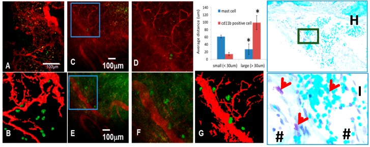
Although most fractures heal, critical defects in bone fail due to aberrant differentiation of mesenchymal stem cells towards fibrosis rather than osteogenesis. While conventional bioengineering solutions to this problem have focused on enhancing angiogenesis, which is required for bone formation, recent studies have shown that fibrotic non-unions are associated with arteriogenesis in the center of the defect and accumulation of mast cells around large blood vessels. Recently, recombinant parathyroid hormone (rPTH; teriparatide; Forteo) therapy have shown to have anti-fibrotic effects on non-unions and critical bone defects due to inhibition of arteriogenesis and mast cell numbers within the healing bone. As this new direction holds great promise towards a solution for significant clinical hurdles in craniofacial reconstruction and limb salvage procedures, this work reviews the current state of the field, and provides insights as to how teriparatide therapy could be used as an adjuvant for healing critical defects in bone. Finally, as teriparatide therapy is contraindicated in the setting of cancer, which constitutes a large subset of these patients, we describe early findings of adjuvant therapies that may present future promise by directly inhibiting arteriogenesis and mast cell accumulation at the defect site.

Keywords: arteriogenesis; critical bone defect; fibrosis; mast cells; osteogenesis; recombinant parathyroid hormone (rPTH; teriparatide; Forteo).


Conflict of interest statement

The authors declare no conflict of interest.

Figures

Figure 1
Figure 1
Mast cells accumulate proximal to large vessels in fibrotic tissue of healing critical defects. The murine chronic cranial defect window model was utilized for in vivo multiphoton laser scanning microscopy (MPLSM) to assess the temporal-spatial relationship of arteriogenesis and mast cell accumulation during critical bone defect healing. MPLSM was performed on mice (n = 5) with cranial defect windows following administration of i.v. Texas red dextran (TRD) and APC-conjugated anti-CD11b or FITC conjugated anti-Mcpt5 antibodies. (A) Representative 10× field of the CD11b+ cells (green) and vasculature (red) with a 3D reconstruction (B), are shown to illustrate that monocytes and macrophages primarily exist proximal to small vessels within the critical defect after three weeks of healing; In contrast, few Mcpt5+ mast cells are found near small vessels ((C); boxed region is (D)); but appear in immediate proximity to large vessels ((E); boxed region is (F) and 3D reconstructed image is (G)); MATLAB quantification of the distance of the labeled cells from small and large vessels is presented as mean +/− SD (* p < 0.05 vs. small vessels). Histologic confirmation of these findings is provided by toluidine blue staining of the calvaria tissue presented at 5× (H); and boxed region at 20× (I); in which the granulated mast cells (red arrows) stain purple in immediate proximity to large blood vessels (#).
Figure 2
Figure 2
Schematic Model of Scarful vs. Scarless Healing and the Adjuvant Effects of rPTH Therapy. An emerging model to explain the fundamental basis of a non-healing critical defect in bone posits that there is a temporal-spatial competition between coupled small vessel angiogenesis-osteogenesis at the leading edge of the healing defect, and large vessel arteriogenesis-fibrosis within the defect. In the first two weeks after injury small vessel (<10 mm) angiogenesis facilitates rapid defect filling (scarless healing) from the leading edge (~0.02 mm/day) via proliferating/migrating bone forming osteoblasts (green). Simultaneously, large vessel (>30 mm) arteriogenesis occurs with the appearance of pro-fibrotic mast cells (dark blue) in the center of the defect, which completely inhibits osteoblastic defect filling (0 mm/day) thereafter, resulting in “scarful healing” of the critical defect. Moreover, rPTH therapy facilitates critical defect healing by: (1) its well-known anabolic effects on osteogenic cells to increase defect filling at the healing front; (2) coupled osteogenic cell-induced small vessel angiogenesis at the healing front; and (3) inhibition of large vessel arteriogenesis/fibrosis within the defect, resulting in scarless healing and ultimate wound closure.

References

    1. Reizner W., Hunter J.G., O’Malley N.T., Southgate R.D., Schwarz E.M., Kates S.L. A systematic review of animal models for staphylococcus aureus osteomyelitis. Eur. Cell Mater. 2014;27:196–212. - PMC - PubMed
    1. Harris J.S., Bemenderfer T.B., Wessel A.R., Kacena M.A. A review of mouse critical size defect models in weight bearing bones. Bone. 2013;55:241–247. doi: 10.1016/j.bone.2013.02.002. - DOI - PMC - PubMed
    1. Marsell R., Einhorn T.A. The biology of fracture healing. Injury. 2011;42:551–555. doi: 10.1016/j.injury.2011.03.031. - DOI - PMC - PubMed
    1. Carano R.A., Filvaroff E.H. Angiogenesis and bone repair. Drug Discov. Today. 2003;8:980–989. doi: 10.1016/S1359-6446(03)02866-6. - DOI - PubMed
    1. Kanczler J.M., Oreffo R.O. Osteogenesis and angiogenesis: The potential for engineering bone. Eur. Cell Mater. 2008;15:100–114. - PubMed

LinkOut - more resources