. 2016 Mar 1;2(1):1-9.
doi: 10.1007/s40883-016-0008-5. Epub 2016 Jan 25.

Multiscale Poly-(ϵ-caprolactone) Scaffold Mimicking Nonlinearity in Tendon Tissue Mechanics

Affiliations

Multiscale Poly-(ϵ-caprolactone) Scaffold Mimicking Nonlinearity in Tendon Tissue Mechanics

Brittany L Banik et al. Regen Eng Transl Med. .

Abstract

Regenerative medicine plays a critical role in the future of medicine. However, challenges remain to balance stem cells, biomaterial scaffolds, and biochemical factors to create successful and effective scaffold designs. This project analyzes scaffold architecture with respect to mechanical capability and preliminary mesenchymal stem cell response for tendon regeneration. An electrospun fiber scaffold with tailorable properties based on a "Chinese-fingertrap" design is presented. The unique criss-crossed fiber structures demonstrate non-linear mechanical response similar to that observed in native tendon. Mechanical testing revealed that optimizing the fiber orientation resulted in the characteristic "S"-shaped curve, demonstrating a toe region and linear elastic region. This project has promising research potential across various disciplines: vascular engineering, nerve regeneration, and ligament and tendon tissue engineering.

Keywords: bioinstructive scaffold; electrospinning; poly-(ϵ-caprolactone); regenerative medicine; tendon.


Figures

Fig. 1
Fig. 1
Electrospinning set-up for tendon scaffold synthesis a) The collecting target is a rod rotating twice counterclockwise and then switching directions and rotating twice clockwise, b) Device mechanism used to rotate the collecting rod target
Fig. 2
Fig. 2
Proposed tendon scaffold a) Cartoon of the tendon scaffold illustrating the Chinese-fingertrap design—when the scaffold is pulled on the ends, a change in length is associated with a change in radius, b) Image of final electrospun PCL tendon scaffold
Fig. 3
Fig. 3
Histograms and SEM images representing fiber alignment on scaffolds produced at various speeds: 184 rpm, 216 rpm, 264 rpm, and 328 rpm and corresponding fiber diameters
Fig. 4
Fig. 4
Mechanical suitability of scaffolds a) Natural tendon stress-strain curve. The `toe region' from 0% – 3% is noteworthy. This represents the point at which the crimped collagen fibers are elongating and stretching until they are fully extended and reach the linear section of the curve Reprinted from Journal of Biomechanics 39(9), J. H.-C. Wang, “Mechanobiology of Tendon,” 1563–1582, 2006, with permission from Elsevier b) Representative stress-strain curve for scaffolds. The toe region falls from 0% – 2.7% with an elastic modulus of 35.8 MPa for the represented scaffold, which is a scaffold spun at 264 rpm
Fig. 5
Fig. 5
Tensile testing for scaffold mechanics a) Scaffold moduli based on rotational speed, b) Scaffold toe region based on rotational speed, c) Tendon scaffold mounted on 3D-printed stubs in tensile testing machine prior to test (left) and stretched scaffold during tensile testing experiment (right) (* p < 0.05, ** p < 0.01, *** p < 0.001)
Fig. 6
Fig. 6
Preliminary cell work a) hMSCs exhibit early contact guidance phenomenon, b) hMSCs adhere and align along fibers forming a cell sheet in the direction of the bulk scaffold at a later timepoint (red:actin:Phalloidin, blue:nuclei:DAPI); arrows indicate direction of bulk scaffold
Fig. 7
Fig. 7
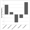
Preliminary gene expression analysis: Reverse-transcription PCR (RT-PCR) data presented as average fold regulation, cells seeded on glass coverslip served as the control surface, scaffold fiber diameter = 271 nm

References

    1. Maffulli N, Renstrom P, Leadbetter WB, editors. Tendon Injuries: Basic Science and Clinical Medicine. Springer; 2005.
    1. Chen J, Xu J, Wang A, Zheng M. Scaffolds for Tendon and Ligament Repair: Review of the Efficacy of Commercial Products. Expert Rev Med Devices. 2009;6:61–73. doi:10.1586/17434440.6.1.61. - PubMed
    1. Martin B, Burr DB, Sharkey NA. Mechanical Properties of Ligament and Tendon. In: Martin B, Burr DB, Sharkey NA, editors. Skelet. Tissue Mech. Springer; New York, NY: 1998. pp. 309–47.
    1. Lim JJ, Temenoff JS. Tendon and Ligament Tissue Engineering: Restoring Tendon/Ligament and Its Interfaces. In: Meyer U, Meyer T, Handschel J, Wiesmann HP, editors. Fundam. Tissue Eng. Regen. Med. Springer; 2009. pp. 255–69. doi:10.1007/978-3-540-77755-7.
    1. Galatz LM, Gerstenfeld L, Heber-Katz E, Rodeo SA. Tendon Regeneration and Scar Formation: The Concept of Scarless Healing. J Orthop Res. 2015;33:823–31. doi:10.1002/jor.22853. - PMC - PubMed