Review
. 2016 May 3:15:70.
doi: 10.1186/s12934-016-0468-9.

Display of recombinant proteins at the surface of lactic acid bacteria: strategies and applications

Affiliations
Review

Display of recombinant proteins at the surface of lactic acid bacteria: strategies and applications

C Michon et al. Microb Cell Fact. .

Abstract

Lactic acid bacteria (LAB) are promising vectors of choice to deliver active molecules to mucosal tissues. They are recognized as safe by the World Health Organization and some strains have probiotic properties. The wide range of potential applications of LAB-driven mucosal delivery includes control of inflammatory bowel disease, vaccine delivery, and management of auto-immune diseases. Because of this potential, strategies for the display of proteins at the surface of LAB are gaining interest. To display a protein at the surface of LAB, a signal peptide and an anchor domain are necessary. The recombinant protein can be attached to the membrane layer, using a transmembrane anchor or a lipoprotein-anchor, or to the cell wall, by a covalent link using sortase mediated anchoring via the LPXTG motif, or by non-covalent liaisons employing binding domains such as LysM or WxL. Both the stability and functionality of the displayed proteins will be affected by the kind of anchor used. The most commonly surfaced exposed recombinant proteins produced in LAB are antigens and antibodies and the most commonly used LAB are lactococci and lactobacilli. Although it is not necessarily so that surface-display is the preferred localization in all cases, it has been shown that for certain applications, such as delivery of the human papillomavirus E7 antigen, surface-display elicits better biological responses, compared to cytosolic expression or secretion. Recent developments include the display of peptides and proteins targeting host cell receptors, for the purpose of enhancing the interactions between LAB and host. Surface-display technologies have other potential applications, such as degradation of biomass, which is of importance for some potential industrial applications of LAB.

Keywords: Genetic engineering; Lactic acid bacteria; Surface display.


Figures
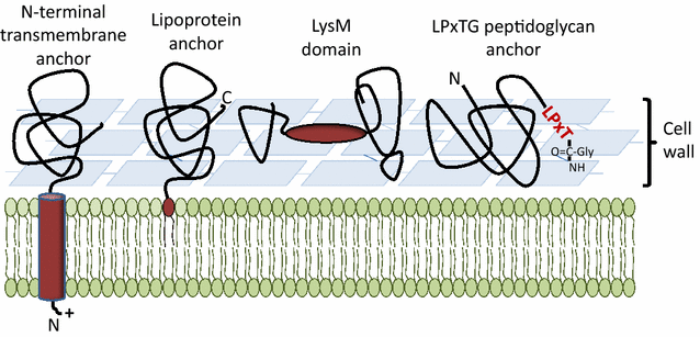
Fig. 1
Fig. 1
Methods for protein display in lactobacilli. A schematic view of the most exploited anchoring methods that are based on covalent or non-covalent interactions with components of the cell membrane or the cell wall. Dark red shows anchor domains/motifs, which are coupled to the to-be-displayed protein. The various routes for display are discussed in the main text. Additional variants based on using a binding domain (i.e. similar to the LysM domain-based strategy) are also discussed in the text. Note that the LysM domain may have various positions relative to the to-be expressed protein; see text
Fig. 2
Fig. 2
Covalent anchoring to the lactobacillal cell wall. The picture shows a schematic overview of the expression cassette for cell wall anchoring of a protein of interest, using an anchoring sequence derived from the Lp_2578 protein of L. plantarum. The cassette is translationally fused to the inducible PsppA promoter and all parts are easily interchangeable using introduced restriction sites (NdeI, SalI, MluI and multiple cloning site, MCS). The primary gene product comprises a signal peptide (indigo), which in this example is derived from Lp_2578, but which could be any peptide from a signal peptide library [23]. The predicted signal peptide cleavage site is indicated by an arrow. The protein of interest is inserted between SalI and MluI restriction sites that were engineered into the vector for this purpose (i.e. two two-residue linker sequences, in green). The protein of interest is C-terminally fuses to a C-terminal fragment of Lp_2578 including the LPxTG anchoring domain consisting of the LPxTG motif (red; the consensus sequence in L. plantarum is LPQTxE [148]), followed by a highly hydrophobic stretch (black) and positively charged C-terminal arginine residues (blue). The length of the Lp_2578 linker may be varied and so far three variants have been constructed, all of which were shown to work [41]. Full length N-terminally processed and cell-wall anchored Lp_2578 is 647 residues of which the last 194 residues are a region of low complexity. The three available linkers comprise 128, 194 and 644 residues, corresponding to a truncated low-complexity region, the complete low complexity region, and almost the complete protein, respectively
Fig. 3
Fig. 3
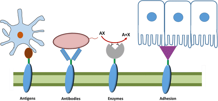
Global view of applications of protein display at surface of LAB. Antigen display at surface of LAB can be used for various goals as: increasing the immune response (antigen display; adhesive protein display; antibodies display), metabolic engineering (enzyme display) or provide a passive immune response (antibodies display)
Fig. 4
Fig. 4
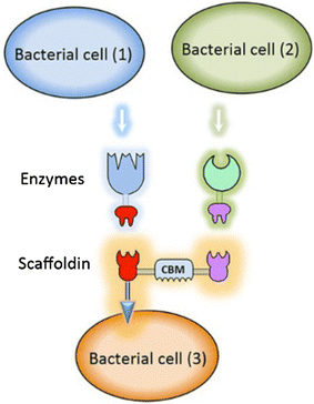
Display of a mini-cellulosome. The picture, taken from Morais et al. [139], BfB, shows how a consortium of recombinant lactobacilli may be used to create LAB displaying a mini-cellulosome. Two strains produce one secreted glycoside hydrolase each; the enzymes are fused to dockerin domains. A third strain produces a scaffoldin that is anchored to the cell surface. The secreted glycoside hydrolases will dock onto the scaffoldin through their dockerin domains, thus forming a mini-cellulosome

References

    1. Gareau M, Sherman P, Walker W. Probiotics and the gut microbiota in intestinal health and disease. Nat Rev Gastroenterol Hepathology. 2010;7:503–514. doi: 10.1038/nrgastro.2010.117. - DOI - PMC - PubMed
    1. Vesa T, Pochart P, Marteau P. Pharmacokinetics of Lactobacillus plantarum NCIMB 8826, Lactobacillus fermentum KLD, and Lactococcus lactis MG 1363 in the human gastrointestinal tract. Aliment Pharmacol Ther. 2000;14:823–828. doi: 10.1046/j.1365-2036.2000.00763.x. - DOI - PubMed
    1. Bermúdez-Humarán LG, Aubry C, Motta J-P, Deraison C, Steidler L, Vergnolle N, Chatel J-M, Langella P. Engineering lactococci and lactobacilli for human health. Curr Opin Microbiol. 2013;16:278–283. doi: 10.1016/j.mib.2013.06.002. - DOI - PubMed
    1. Motta J-P, Bermúdez-Humarán LG, Deraison C, Martin L, Rolland C, Rousset P, Boue J, Dietrich G, Chapman K, Kharrat P, Vinel J-P, Alric L, Mas E, Sallenave J-M, Langella P, Vergnolle N. Food-grade bacteria expressing elafin protect against inflammation and restore colon homeostasis. Sci Transl Med. 2012;4:158ra144. doi: 10.1126/scitranslmed.3004212. - DOI - PubMed
    1. Galipeau HJ, Wiepjes M, Motta J-P, Schulz JD, Jury J, Natividad JM, Pinto-Sanchez I, Sinclair D, Rousset P, Martin-Rosique R, Bermudez-Humaran L, Leroux JC, Murray JA, Smecuol E, Bai JC, Vergnolle N, Langella P, Verdu EF. Novel role of the serine protease inhibitor elafin in gluten-related disorders. Am J Gastroenterol. 2013;2014:1–9. - PMC - PubMed

Publication types

MeSH terms