. 2017 Jan;10(1):91-103.
doi: 10.1038/mi.2016.45. Epub 2016 May 4.

Core 1- and 3-derived O-glycans collectively maintain the colonic mucus barrier and protect against spontaneous colitis in mice

Affiliations

Core 1- and 3-derived O-glycans collectively maintain the colonic mucus barrier and protect against spontaneous colitis in mice

K Bergstrom et al. Mucosal Immunol. 2017 Jan.

Abstract

Core 1- and 3-derived mucin-type O-glycans are primary components of the mucus layer in the colon. Reduced mucus thickness and impaired O-glycosylation are observed in human ulcerative colitis. However, how both types of O-glycans maintain mucus barrier function in the colon is unclear. We found that C1galt1 expression, which synthesizes core 1 O-glycans, was detected throughout the colon, whereas C3GnT, which controls core 3 O-glycan formation, was most highly expressed in the proximal colon. Consistent with this, mice lacking intestinal core 1-derived O-glycans (IEC C1galt1-/-) developed spontaneous colitis primarily in the distal colon, whereas mice lacking both intestinal core 1- and 3-derived O-glycans (DKO) developed spontaneous colitis in both the distal and proximal colon. DKO mice showed an early onset and more severe colitis than IEC C1galt1-/- mice. Antibiotic treatment restored the mucus layer and attenuated colitis in DKO mice. Mucins from DKO mice were more susceptible to proteolysis than wild-type mucins. This study indicates that core 1- and 3-derived O-glycans collectively contribute to the mucus barrier by protecting it from bacterial protease degradation and suggests new therapeutic targets to promote mucus barrier function in colitis patients.


Conflict of interest statement

The authors declared no conflict of interest.

Figures

Figure 1
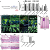
Figure 1. DKO mice have early onset and more severe colitis in the distal colon
(a) Representative images of H&E stained distal colons. P12 = postnatal day 12. (b) Histologic colitis score. (c) Representative AB staining of Carnoy’s-fixed colonic sections. (d) Quantification of mucus thickness vs. WT mice. Error bars = SEM. (e) Representative images of Tn antigen immunohistochemistry (IHC) (brown) in colon sections. Arrow: top of mucus layer. (f) Quantification of Tn+ cells from mice in (e). (g) Representative dual staining for Muc2 (IF, green) and bacteria (FISH, red), the latter using the universal EUB338 probe, on Carnoy’s-fixed colonic sections. Inset, magnified image of boxed region. Arrowheads, bacteria. (h) Quantitation of serum FITC-Dextran, 4 kDa (FD4) 4 h after oral administration. Bars show average fold changes relative to WT mice. Data are representative of >2 independent studies with n = 4 – 6 mice/group. For (b), (d), and (h) *P < 0.05 vs. WT.
Figure 2
Figure 2. DKO mice exhibit defective mucus and spontaneous colitis in the proximal colon
(a) Gene expression analysis of enriched colonic crypt cells by RT-qPCR. (b) Representative IHC for Tn antigen (brown). Inset, magnified image of boxed regions. Arrowheads: red, Tn goblet cell; black, Tn+ goblet cell. (c) Quantification Tn+ goblet cells. Error bars = SEM. (d) Representative IF staining for Muc2 on Carnoy’s-fixed sections. Arrows: White, mucus layer; yellow, luminal food particle. (e) Quantification of mucus thickness vs. WT mice. (f) Representative H&E staining of proximal colonic sections. (g) Histologic colitis score. Data are representative of 2 independent studies, n = 4 – 6/group; WT mice are littermate controls. *P < 0.05, student’s t test for (a), (c), (e) and 1 way ANOVA with Bonferonni post-test for (g).
Figure 3
Figure 3. Deficiency of core 1- and 3-derived O-glycans impairs mucus layer and causes colitis in adult mice
(a) Generation of inducible DKO mice. (b) Experimental design. (c) H&E staining of carnoy’s fixed distal colon sections. (d) Histologic colitis score. (e) Representative AB staining on Carnoy’s fixed colon sections. (f) Quantification of mucus thickness vs. WT mice (from Muc2 stain). (g) Dual IF for Muc2 (green) and Tn antigen (red); dual Muc2+Tn+ cells are yellow (white arrow). Arrowheads: white, Tn portion of mucus; red, Tn+ portion of mucus. (h) Quantification of dual Muc2+Tn+ cells. For all bar graphs, error bars = SEM. Results are representative of 2 independent experiments, n = 4 – 6 mice/group/time point. TM-treated WT littermates are experimental controls. *P < 0.05 vs. TM-treated WT mice, 1 way ANOVA with Bonferroni post test.
Figure 4
Figure 4. Microbial depletion rescues defective mucus layer function and ameliorates colitis in DKO mice
(a) Experimental design. (b) qPCR of bacterial universal and group-specific 16S RNA gene from fecal gDNA.. (c) Representative dual Muc2/EUB33 staining. Arrow, mucus layer. (d) Composite AgPAGE analysis of semipurified murine colonic mucin, visualized by PAS staining (magenta). Arrow, mucin band in DKO. (e) IF staining for the lectin UEA-1. Arrow, mucus layer. (f) Representative H&E staining of distal colon sections. Right: histologic damage score. Results are representative of 2 independent experiments, n = 4 mice/group. WT mice are experimental controls. *P < 0.05 and **P < 0.01 vs. vehicle-treated WT, 1 way ANOVA with Bonferroni post-test.
Figure 5
Figure 5. Loss of the mucus barrier in DKO deficient mice is dependent upon the microbiota but not inflammation
(a) Experimental design for short-term microbial depletion. (b) qPCR of total bacterial universal 16S RNA gene. (c) qPCR of bacterial 16S RNA gene specific for Bacteroidales order. (d) Representative FISH using EUB338 to stain bacteria (red). Right: Relative quantification of FISH signal in lumen. (e) Representative H&E of distal colon tissues. Right; histologic damage score. (f) Dual IF for Muc2 (green) and Tn antigen (red) on Carnoy’s-fixed colonic sections. Arrows, dual Muc2+Tn+ mucus layer. (g) Composite AgPAGE analysis of semipurified murine colonic mucin, visualized by PAS staining (magenta). Arrow, DKO Muc2 mucin band. CB, Coomassie Blue loading control. Results are representative of 3 independent experiments, 3 – 6 mice/group. Non-treated WT or DKO are experimental controls. *P < 0.05, student’s t test.
Figure 6
Figure 6. O-glycans protect Muc2 from bacterial protease-mediated degradation
(a) Dual Muc2/FISH staining. Note: images are from mice on a gel-food diet, which allowed preservation of broken mucus in untreated DKO mice. Arrows, bacteria. (b) PAS-stained composite AgPAGE gel of mucin derived from streptomycin-treated mice. CB, Coomassie Blue loading control. (c) Western blot for Muc2 protein (αMuc2-C3) and Fucosylated Muc2 (UEA1). (d) Densitometry of mature and immature Muc2 bands in WT and DKO mice. Error bars, SEM. Degradation studies are representative of 3 separate experiments, the third with independently derived mucin samples. *P < 0.05 vs. 0 mU, student’s t test.

References

    1. Ford AC, Moayyedi P, Hanauer SB. Ulcerative colitis. BMJ. 2013;346 - PubMed
    1. Strober W, Fuss I, Mannon P. The fundamental basis of inflammatory bowel disease. J Clin Invest. 2007;117:514–521. - PMC - PubMed
    1. Johansson ME, Sjovall H, Hansson GC. The gastrointestinal mucus system in health and disease. Nat Rev Gastroenterol Hepatol. 2013;10:552–361. - PMC - PubMed
    1. Ley RE, Peterson DA, Gordon JI. Ecological and evolutionary forces shaping microbial diversity in the human intestine. Cell. 2006;124:837–848. - PubMed
    1. Pelaseyed T, et al. The mucus and mucins of the goblet cells and enterocytes provide the first defense line of the gastrointestinal tract and interact with the immune system. Immunol Rev. 2014;260:8–20. - PMC - PubMed

Publication types

MeSH terms

Substances