

3T MRI investigation of cardiac left ventricular structure and function in a UK population: The tayside screening for the prevention of cardiac events (TASCFORCE) study
- PMID: 27143317
- PMCID: PMC5082537
- DOI: 10.1002/jmri.25267
3T MRI investigation of cardiac left ventricular structure and function in a UK population: The tayside screening for the prevention of cardiac events (TASCFORCE) study
Abstract
Purpose: To scan a volunteer population using 3.0T magnetic resonance imaging (MRI). MRI of the left ventricular (LV) structure and function in healthy volunteers has been reported extensively at 1.5T.
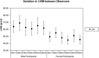
Materials and methods: A population of 1528 volunteers was scanned. A standardized approach was taken to acquire steady-state free precession (SSFP) LV data in the short-axis plane, and images were quantified using commercial software. Six observers undertook the segmentation analysis.
Results: Mean values (±standard deviation, SD) were: ejection fraction (EF) = 69 ± 6%, end diastolic volume index (EDVI) = 71 ± 13 ml/m2 , end systolic volume index (ESVI) = 22 ± 7 ml/m2 , stroke volume index (SVI) = 49 ± 8 ml/m2 , and LV mass index (LVMI) = 55 ± 12 g/m2 . The mean EF was slightly larger for females (69%) than for males (68%), but all other variables were smaller for females (EDVI 68v77 ml/m2 , ESVI 21v25 ml/m2 , SVI 46v52 ml/m2 , LVMI 49v64 g/m2 , all P < 0.05). The mean LV volume data mostly decreased with each age decade (EDVI males: -2.9 ± 1.3 ml/m2 , females: -3.1 ± 0.8 ml/m2 ; ESVI males: -1.3 ± 0.7 ml/m2 , females: -1.7 ± 0.5 ml/m2 ; SVI males: -1.7 ± 0.9 ml/m2 , females: -1.4 ± 0.6 ml/m2 ; LVMI males: -1.6 ± 1.1 g/m2 , females: -0.2 ± 0.6 g/m2 ) but the mean EF was virtually stable in males (0.6 ± 0.6%) and rose slightly in females (1.2 ± 0.5%) with age.
Conclusion: LV reference ranges are provided in this population-based MR study at 3.0T. The variables are similar to those described at 1.5T, including variations with age and gender. These data may help to support future population-based MR research studies that involve the use of 3.0T MRI scanners. J. Magn. Reson. Imaging 2016;44:1186-1196.
Keywords: 3.0T; MRI; cardiac; left ventricle; population.
© 2016 The Authors Journal of Magnetic Resonance Imaging published by Wiley Periodicals, Inc. on behalf of International Society for Magnetic Resonance in Medicine.
Figures

References
-
- Alfakih K, Plein S, Thiele H, Jones T, Ridgway JP, Sivananthan MU. Normal human left and right ventricular dimensions for MRI as assessed by turbo gradient echo and steady‐state free precession imaging sequences. J Magn Reson Imaging JMRI 2003;17:323–329. - PubMed
-
- Hudsmith LE, Petersen SE, Francis JM, Robson MD, Neubauer S. Normal human left and right ventricular and left atrial dimensions using steady state free precession magnetic resonance imaging. J Cardiovasc Magn Reson 2005;7:775–782. - PubMed
-
- Maciera AM, Prasad SK, Khan M, Pennell DJ. Normalized Left ventricular systolic and diastolic function by steady state free precession cardiovascular magnetic resonance. J Cardiovasc Magn Reson 2006;8:417–426. - PubMed
-
- Chung AK, Das SR, Leonard D, et al. Women have higher left ventricular ejection fractions than men independent of differences in left ventricular volume: the Dallas Heart Study. Circulation 2006;113:1597–1604. - PubMed
Publication types
MeSH terms
Grants and funding
LinkOut - more resources
Full Text Sources
Other Literature Sources
Medical
