. 2016 May 3;5(5):e003474.
doi: 10.1161/JAHA.116.003474.

Local Application of Leptin Antagonist Attenuates Angiotensin II-Induced Ascending Aortic Aneurysm and Cardiac Remodeling

Affiliations

Local Application of Leptin Antagonist Attenuates Angiotensin II-Induced Ascending Aortic Aneurysm and Cardiac Remodeling

Danny Ben-Zvi et al. J Am Heart Assoc. .

Abstract

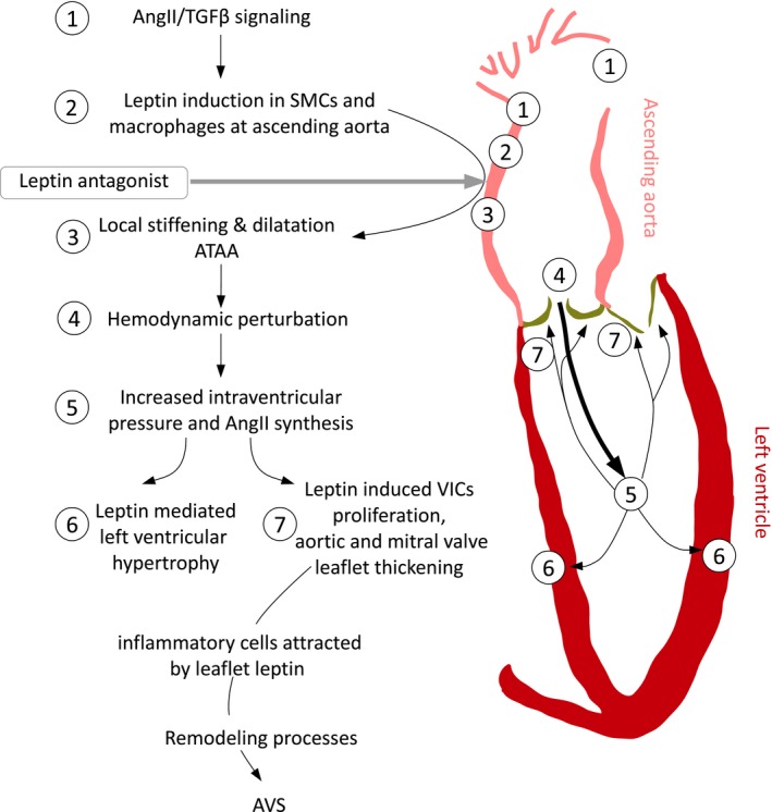
Background: Ascending thoracic aortic aneurysm (ATAA) is driven by angiotensin II (AngII) and contributes to the development of left ventricular (LV) remodeling through aortoventricular coupling. We previously showed that locally available leptin augments AngII-induced abdominal aortic aneurysms in apolipoprotein E-deficient mice. We hypothesized that locally synthesized leptin mediates AngII-induced ATAA.

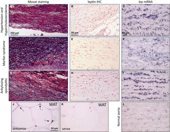
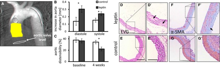
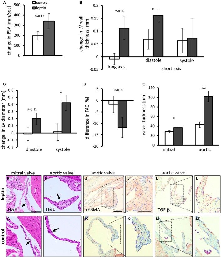
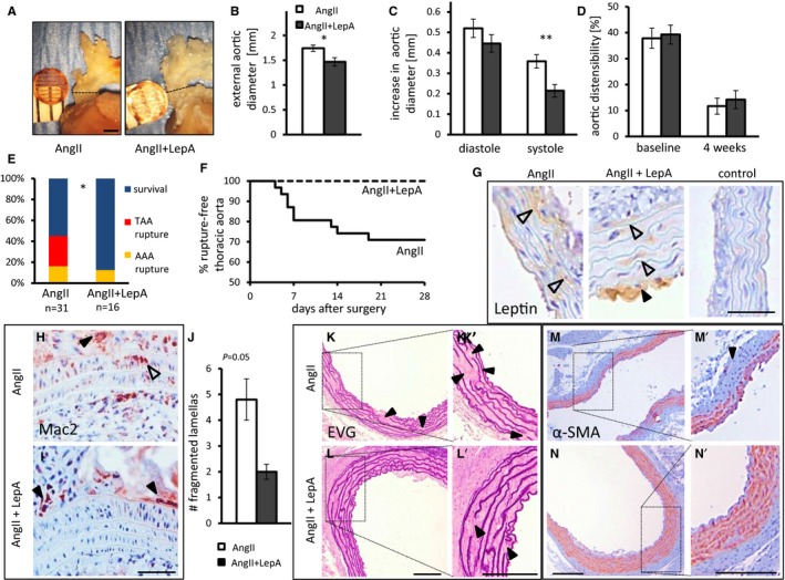
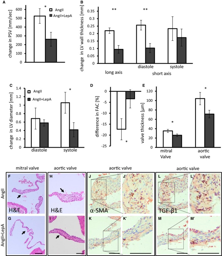
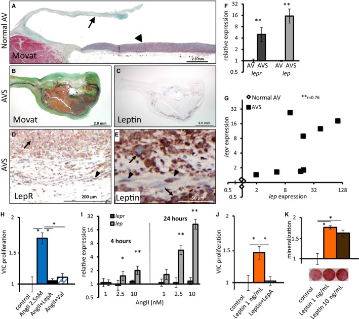
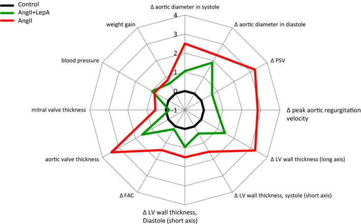
Methods and results: Following demonstration of leptin synthesis in samples of human ATAA associated with different etiologies, we modeled in situ leptin expression in apolipoprotein E-deficient mice by applying exogenous leptin on the surface of the ascending aorta. This treatment resulted in local aortic stiffening and dilation, LV hypertrophy, and thickening of aortic/mitral valve leaflets. Similar results were obtained in an AngII-infusion ATAA mouse model. To test the dependence of AngII-induced aortic and LV remodeling on leptin activity, a leptin antagonist was applied to the ascending aorta in AngII-infused mice. Locally applied single low-dose leptin antagonist moderated AngII-induced ascending aortic dilation and protected mice from ATAA rupture. Furthermore, LV hypertrophy was attenuated and thickening of aortic valve leaflets was moderated. Last, analysis of human aortic valve stenosis leaflets revealed de novo leptin synthesis, whereas exogenous leptin stimulated proliferation and promoted mineralization of human valve interstitial cells in culture.

Conclusions: AngII-induced ATAA is mediated by locally synthesized leptin. Aortoventricular hemodynamic coupling drives LV hypertrophy and promotes early aortic valve lesions, possibly mediated by valvular in situ leptin synthesis. Clinical implementation of local leptin antagonist therapy may attenuate AngII-induced ATAA and moderate related LV hypertrophy and pre-aortic valve stenosis lesions.

Clinical trial registration: URL: https://www.clinicaltrials.gov/. Unique identifier: NCT00449306.

Keywords: angiotensin II; aortic aneurysm; aortic valve stenosis; left ventricular hypertrophy; leptin; leptin antagonist; vascular remodeling.


Figures

Figure 1
Figure 1
Analysis of surgical ATAA samples collected from patients with a variety of diseases underlying ATAA. A through C, Hypertension and hypercholesterolemia. D through F, Marfan syndrome. G through I, Ankylosing spondylitis. A, D, and G, Diffuse elastic fiber fragmentation with glycosaminoglycan deposition (bluish‐green), matrix clearing, and media degeneration, Movat's pentachrome. Scale bar=250 μm. B, E, and H, Leptin immunostaining in medial smooth muscle cells. Scale bar=50 μm. C, F, I, and L, The lep mRNA (black staining) is present in diseased aortas but is scarcely present in normal aorta. Scale bar=10 μm. J and K, Positive (antisense) (J) and negative (sense) (K) control for lep mRNA. In situ hybridization in human WAT. Scale bar=10 μm. ATAA indicates ascending thoracic aortic aneurysm; IHC, immunohistochemistry; WAT, white adipose tissue.
Figure 2
Figure 2
The effects of locally applied leptin on the ascending aorta. A, Human arch arteriogram depicting mouse aortic anatomy. Note the location of leptin slow‐release film (yellow) in relation to the aortic valve level (white dashed lines). B, Increase in maximal aortic diameter from baseline following treatment with leptin in diastole and systole (P=0.02 and P=0.045, respectively). Baseline aortic diameter was 1.3 mm. C, Percentage of aortic wall distensibility assessed at baseline and 4 weeks after surgery (P=0.01; n=10 to 11). Control mice in white bars, leptin treated in gray bars. *P<0.05. D through G, Histologic cross‐sections of the ascending aortic at the location of leptin application, as shown in (A). Elastic Van Gieson (EVG) staining shows fragmentations of elastic lamellae (arrows) in leptin treated (D) and control mice (E). Depletion of α‐smooth muscle actin (α‐SMA) immunostaining (arrow) in leptin treated (F) and control mice (G). Scale bar=100 μm, 200 μm for (D′ and E′).
Figure 3
Figure 3
Leptin applied at the ascending aorta induces remodeling of LV wall and valve leaflets. A, Leptin‐induced increase in PSV measured at the aortic valve. P=0.17. Mean baseline PSV was 1150 mm/s. B, Leptin induced an increase in LV wall thickness, as measured in the long‐axis view (P=0.06), and in diastole, as measured in the short‐axis view (P=0.045). Mean LV wall thickness at baseline was 0.83 mm at anterior and posterior walls and 0.55 mm at the septum. C, Increase in LV diameter following treatment with leptin in diastole (P=0.11) and in systole (P=0.04). Mean LV diameter at baseline was 2.2 mm. D, Difference in FAC following treatment with leptin (P=0.09). Mean FAC at baseline was 63%. E, Thickness of mitral and aortic valve leaflets following leptin treatment (P=0.02 and P=0.006, respectively). A through E, n=9 to 11. Control mice shown in white bars; leptin‐treated mice shown in dark gray bars. F and G, H&E staining of mitral valve from leptin‐treated (F) and control (G) mice. Arrows point to the valve. Scale bar=100 μm. H and I, H&E staining of aortic valve from leptin‐treated (H) and control (I) mice. Arrows point to the valve. J and K, α‐SMA staining in aortic valves of leptin‐treated mice (J) and controls (K). L and M, TGF‐β1 expression in aortic valves of leptin‐treated mice (L) and controls (M). Scale bar: Low magnification=50 μm, enlargements=100 μm. *P<0.05, **P<0.01. α‐SMA indicates α‐smooth muscle cell actin; FAC, fractional area change; H&E, hematoxylin and eosin; LV, left ventricular; PSV, peak systolic velocity; TGF‐β1, transforming growth factor β1.
Figure 4
Figure 4
Local application of LepA at the proximal ascending aorta attenuates the effects of AngII infusion on the aortic wall locally. A and B, External aortic diameter 4 weeks after surgery. n=5, P=0.047. C, Increase in aorta diameter in diastole and systole (P<0.01 in systole). Mean internal aortic diameter was 1.3 mm at baseline. D, Aortic distensibility at baseline and 4 weeks after surgery. C and D, n=12 to 13. E, AngII infusion leads to fatal rupture of TAA or AAA. Surviving mice were euthanized 4 weeks after surgery. Application of LepA abolished TAA rupture (P=0.01, 2‐tailed Fisher exact test). F, A Kaplan–Meier survival curve for mice receiving AngII infusion or AngII cotreated with LepA. G, Leptin was detected in smooth muscle cells (open arrowheads) and atherosclerotic plaques (black arrowhead) but was barely detected in control mice treated with an empty polylactic co–glycolic acid film. Scale bar=50 μm. H and I, Macrophage infiltration in AngII (H) and AngII and LepA‐treated mice (I). Open arrowhead points to infiltrating macrophages, black arrowheads point to macrophages in the adventitia. Scale bar=50 μm. We did not detect macrophages infiltrating the media of LepA‐treated mice (I). J, Number of breaks in aortic elastic lamellas. P=0.05, n=5 to 6. K and L′, Ascending aorta from AngII (K) and AngII and LepA‐treated mice (L) stained by EVG; black arrowheads highlight fragmentation of elastic lamellae. Scale bar=100 μm. M and N′, Immunostaining of α‐SMA in similar location as (K and L′); black arrowhead highlights an absence of α‐SMA. Scale bar=100 μm. *P<0.05, **P<0.01. α‐SMA indicates α‐smooth muscle cell actin; AAA, abdominal aortic aneurysm; AngII, angiotensin II; EVG, elastic Van Gieson; LepA, leptin antagonist; TAA, thoracic aortic aneurysm.
Figure 5
Figure 5
Local application of LepA at the proximal ascending aorta attenuates the effects of AngII infusion on LV remodeling. A, Increase in PSV measured at the aortic valve. P=0.03. AngII‐infused mice shown in white bars; AngII and LepA shown in dark gray bars. Mean PSV at baseline was 1275 mm/s. B, Increase in LV wall thickness as measured by echocardiography in long axis (P<0.01) and in diastole in short axis views (P<0.01). Mean LV wall thickness at baseline was 0.79 mm at anterior and posterior walls and 0.57 mm at the septum. C, Increase in LV diameter in diastole (P=0.047) and systole. Mean LV diameter was 2.3 mm at baseline. D, FAC is reduced in AngII‐infused mice but is not affected in AngII and LepA‐treated mice. P=0.01. Mean FAC was 55% at baseline. E, Mitral and aortic valve leaflets thickness is reduced in AngII and LepA‐treated mice compared with AngII‐infused mice. P=0.03 for both valves. A through E, n=10 to 12. F and G, H&E staining of a mitral valve from AngII (F) and AngII and LepA‐treated mice (G). Arrows point to the valve. H and I, H&E staining of an aortic valve from AngII (H) and AngII and LepA‐treated mice (I). Arrows point to the valve. J and K, α‐SMA is abundant in aortic valves of AngII‐infused mice (J), with relatively weaker expression in AngII and LepA‐treated mice (K). L and M, TGF‐β1 expression is widespread in aortic valves of AngII‐infused mice (L) but is less prevalent in AngII and LepA‐treated mice (M). Scale bar=100 μm (F through M). *P<0.05, **P<0.01. α‐SMA indicates α‐smooth muscle cell actin; AngII, angiotensin II; FAC, fractional area change; H&E, hematoxylin and eosin; LepA, leptin antagonist; LV, left ventricular; PSV, peak systolic velocity; TGF‐β1, transforming growth factor β1.
Figure 6
Figure 6
Leptin is expressed in AVs of AVS patients and induces VIC proliferation and calcification in vitro. A, Movat staining of healthy human AV. Arrowhead points to ascending aorta, arrow to AV. B and C, Movat (B) and leptin IHC (C) of AV collected from an AVS patient. D and E, LepR (D) and leptin (E) IHC from the same valve as in (B and C). Arrows point to macrophage‐like cells; arrowheads point to elongated SMC smooth muscle‐like cells. F, qPCR analysis for lep and lepr expression in AVs. n=3 and n=8 for valves of healthy and AVS patients, respectively. G, Positive correlation between lep and lepr expression in AVs. r 2=0.76 (Spearman correlation), P=0.007. H through K, In vitro primary human VICs. H, AngII increases VIC proliferation. This effect is blocked by the AngII type 1 receptor blocker valsartan and LepA. (P=0.01; n=5–6). I, qPCR analysis for lepr and lep expression in VICs treated with AngII for 4 or 24 hours compared with untreated VICs. P=0.046, P=0.007 (4 hours), P<0.001 (24 hours). Error bars represent standard deviation. J, Leptin increases VICs proliferation (P=0.01), and this effect is blocked by LepA (P=0.01; n=6). K, Mineralization of VICs grown in osteogenic medium is increased by leptin (P<0.05; n=3). *P<0.05, **P<0.01. AngII indicates angiotensin II; AV, aortic valve; AVS, aortic valve stenosis; IHC, immunohistochemistry; LepA, leptin antagonist; LepR, leptin receptor; qPCR, quantitative polymerase chain reaction; Val, valsartan; VIC, valve interstitial cell.
Figure 7
Figure 7
Effects of LepA application at the surface of the ascending aorta on AngII‐induced thoracic aortic aneurysm formation and left ventricular remodeling. A radar plot of quantitative cardiovascular parameters assessed in this study demonstrating the impact of LepA applied on the surface of the ascending aorta in AngII‐infused mice. The distance from center is measured by a Z score calculated from the standard deviation in mice treated with an empty polylactic co–glycolic acid film (black, control). Red line corresponds to the AngII‐infused mice alone, whereas the green line corresponds to the AngII and LepA‐treated mice. Moving clockwise, LepA application attenuates the effects of AngII on ascending aortic diameter, hemodynamics at the LV outlet, LV wall thickness, FAC, and LV valves’ leaflets thickness. There is no effect on blood pressure or weight gain. AngII indicates angiotensin II; FAC, fractional area change; LepA, leptin antagonist; LV, left ventricular; PSV, peak systolic velocity.
Figure 8
Figure 8
A model of a mechanism for AVS initiation through AngII‐induced ATAA, associated with AVC activation. The numerical sequence of events corresponds to findings discussed and shown in this study: Leptin‐mediated ATAA formation, AVC activation underlying left ventricular hypertrophy and aortic valve thickening through leptin‐induced VIC proliferation. AngII indicates angiotensin II; ATAA, ascending thoracic aortic aneurysm; AVC, aortoventricular coupling; AVS, aortic valve stenosis; SMC, smooth muscle cell; TGF‐β, transforming growth factor β; VIC, valve interstitial cell.

Similar articles

Cited by

References

    1. Davies RR, Goldstein LJ, Coady MA, Tittle SL, Rizzo JA, Kopf GS, Elefteriades JA. Yearly rupture or dissection rates for thoracic aortic aneurysms: simple prediction based on size. Ann Thorac Surg. 2002;73:17–27; discussion 27‐8. - PubMed
    1. Danyi P, Elefteriades JA, Jovin IS. Medical therapy of thoracic aortic aneurysms: are we there yet? Circulation. 2011;124:1469–1476. - PubMed
    1. Isselbacher EM. Thoracic and abdominal aortic aneurysms. Circulation. 2005;111:816–828. - PubMed
    1. Lindsay ME, Dietz HC. Lessons on the pathogenesis of aneurysm from heritable conditions. Nature. 2011;473:308–316. - PMC - PubMed
    1. Tadros TM, Klein MD, Shapira OM. Ascending aortic dilatation associated with bicuspid aortic valve: pathophysiology, molecular biology, and clinical implications. Circulation. 2009;119:880–890. - PubMed

MeSH terms

Associated data