. 2016 May 3;7(3):e00270-16.
doi: 10.1128/mBio.00270-16.

Suppression of Antitumor Immune Responses by Human Papillomavirus through Epigenetic Downregulation of CXCL14

Affiliations

Suppression of Antitumor Immune Responses by Human Papillomavirus through Epigenetic Downregulation of CXCL14

Louis Cicchini et al. mBio. .

Abstract

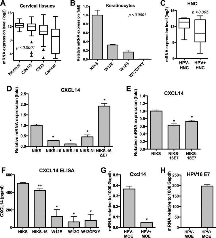
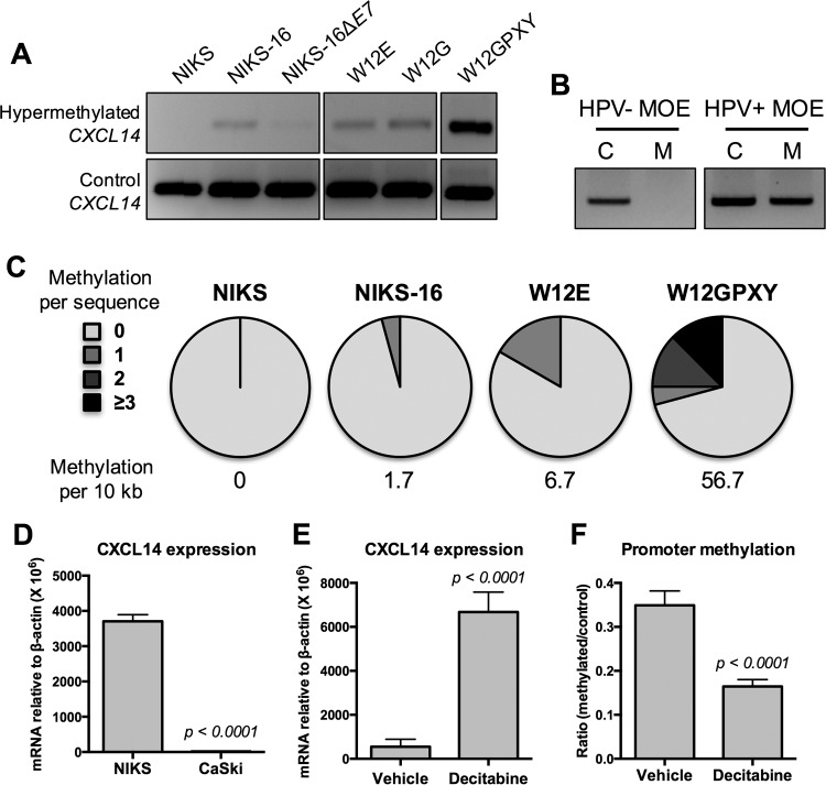
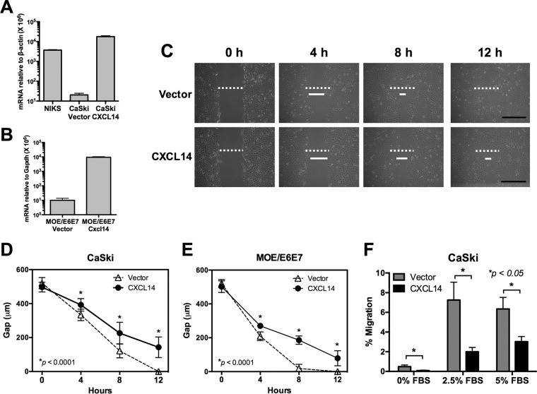
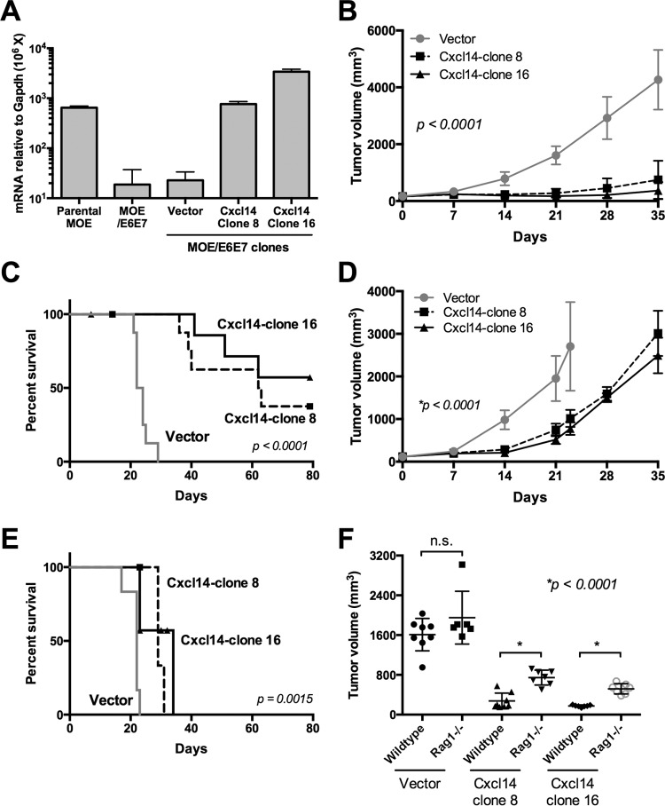
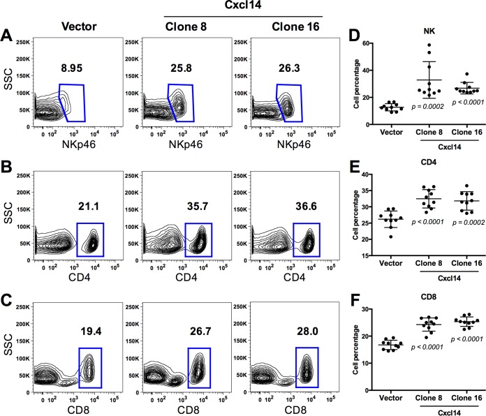
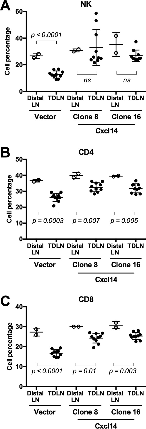
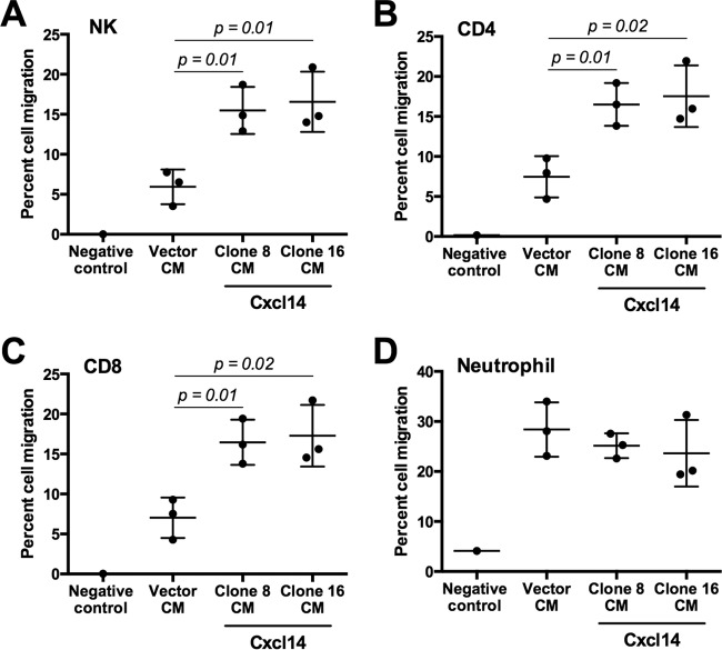
High-risk human papillomaviruses (HPVs) are causally associated with multiple human cancers. Previous studies have shown that the HPV oncoprotein E7 induces immune suppression; however, the underlying mechanisms remain unknown. To understand the mechanisms by which HPV deregulates host immune responses in the tumor microenvironment, we analyzed gene expression changes of all known chemokines and their receptors using our global gene expression data sets from human HPV-positive and -negative head/neck cancer and cervical tissue specimens in different disease stages. We report that, while many proinflammatory chemokines increase expression throughout cancer progression, CXCL14 is dramatically downregulated in HPV-positive cancers. HPV suppression of CXCL14 is dependent on E7 and associated with DNA hypermethylation in the CXCL14 promoter. Using in vivo mouse models, we revealed that restoration of Cxcl14 expression in HPV-positive mouse oropharyngeal carcinoma cells clears tumors in immunocompetent syngeneic mice, but not in Rag1-deficient mice. Further, Cxcl14 reexpression significantly increases natural killer (NK), CD4(+) T, and CD8(+) T cell infiltration into the tumor-draining lymph nodes in vivo In vitro transwell migration assays show that Cxcl14 reexpression induces chemotaxis of NK, CD4(+) T, and CD8(+) T cells. These results suggest that CXCL14 downregulation by HPV plays an important role in suppression of antitumor immune responses. Our findings provide a new mechanistic understanding of virus-induced immune evasion that contributes to cancer progression.

Importance: Human papillomaviruses (HPVs) are causally associated with more than 5% of all human cancers. During decades of cancer progression, HPV persists, evading host surveillance. However, little is known about the immune evasion mechanisms driven by HPV. Here we report that the chemokine CXCL14 is significantly downregulated in HPV-positive head/neck and cervical cancers. Using patient tissue specimens and cultured keratinocytes, we found that CXCL14 downregulation is linked to CXCL14 promoter hypermethylation induced by the HPV oncoprotein E7. Restoration of Cxcl14 expression in HPV-positive cancer cells clears tumors in immunocompetent syngeneic mice, but not in immunodeficient mice. Mice with Cxcl14 reexpression show dramatically increased natural killer and T cells in the tumor-draining lymph nodes. These results suggest that epigenetic downregulation of CXCL14 by HPV plays an important role in suppressing antitumor immune responses. Our findings may offer novel insights to develop preventive and therapeutic tools for restoring antitumor immune responses in HPV-infected individuals.


Figures

FIG 1
FIG 1
CXCL14 expression is downregulated during HPV-associated cancer progression. (A and C) CXCL14 mRNA expression levels were analyzed from global gene expression data sets of cervical tissue samples (A) and HNC tissue samples (C). (A) The 128 cervical tissue samples included samples in different disease stages (normal, n = 24; low-grade lesion, n = 36; high-grade lesion, n = 40; and cancer, n = 28) (4). (C) The 42 HNC tissue samples included 26 HPV-negative (HPV-) HNC and 16 HPV-positive (HPV+) (5) tissue samples. Normalized fluorescence intensities (log2) of gene expression from each group are shown in box-and-whisker plots with Tukey’s method for outliers (black triangles) noted as distinct data points. P values were determined by one-way ANOVA analysis (A) or Student’s t test (C). (B, D, and E) Total RNA was extracted from W12 (B) and NIKS (D and E) cell lines. The expression levels of CXCL14 were measured by RT-qPCR. (F) Secreted CXCL14 was measured by ELISA using culture supernatant from NIKS, NIKS-16, W12E, W12G, and W12GPXY cells. (G and H) Total RNA was extracted from mouse oropharyngeal epithelial (MOE) cell lines, MOE/shPTPN13 (HPV negative) and MOE/E6E7 (HPV positive). The expression levels of murine Cxcl14 mRNA (G) and HPV16 E1^E4 mRNA transcript (H) were measured by RT-qPCR. HPV16 E1^E4 and CXCL14 mRNA copy numbers were calculated using serially diluted standard plasmids and normalized by human β-actin and murine Gapdh mRNA copy numbers. P values were calculated by Student’s t test. Values that are significantly different are indicated by asterisks as follows: *, P < 0.0001; **, P = 0.0002
FIG 2
FIG 2
CXCL14 downregulation in HPV-positive epithelial cells is associated with CXCL14 promoter hypermethylation. (A and B) Genomic DNA was extracted from NIKS, NIKS-16, NIKS-16ΔE7, W12E, W12G, and W12GPXY cells (A) and MOE/shPTPN13 (HPV-negative) and MOE/E6E7 (HPV-positive) cells (B). MSP was performed using specific primers and analyzed in 1.2% agarose gel as described in Text S1 in the supplemental material. (B) MSP products of the control and hypermethylated Cxcl14 promoter are shown in lanes C and M, respectively. (C) Bisulfite PCR products were cloned into the pGEM-T Easy vector and sequenced. (D) CXCL14 expression was measured as described in the legend to Fig. 1. (E and F) CaSki cells were treated with 10 µM decitabine for 6 days or with a vehicle control (H2O) for 6 days. RT-qPCR (E) and qMSP (F) were performed using total RNA and genomic DNA, respectively. CXCL14 mRNA copy numbers were normalized by β-actin mRNA (D and E). (F) Changes in CXCL14 promoter methylation were calculated by using the 2−ΔΔCT method and shown as a fold ratio of methylated signal over total signal. P values were determined by Student’s t test.
FIG 3
FIG 3
CXCL14 expression hinders mobility of HPV-positive cancer cells. (A and B) CaSki and MOE/E6E7 cell lines reexpressing CXCL14 were established using lentiviral transduction of the human CXCL14 and murine Cxcl14 genes, respectively, and validated by RT-qPCR. CXCL14 and Cxcl14 mRNA copy numbers were normalized by human β-actin or murine Gapdh mRNA, respectively. (C to E) In vitro scratch assay was performed with the established CaSki (C and D) and MOE/E6E7 (E) cells. Images were captured 0, 4, 8, and 12 h after the scratch or wound was generated, and the widths of the wound gaps were measured using NIH ImageJ software. Representative data from three replicates of each group are shown. The initial wound gaps (white dashed bar) and representative gaps at the indicated time points (solid white bar) are shown. Bars, 500 µm. (F) Transwell migration assays were performed on CaSki cells reexpressing CXCL14 generated as described above for panel A. The percentage of cells that migrated through the permeable supports is shown, using 0%, 2.5%, and 5% FBS as a generic chemoattractant. P values were calculated by Student’s t test.
FIG 4
FIG 4
Restoration of Cxcl14 expression clears HPV-positive tumors in immunocompetent mice, but not in Rag1-deficient mice. (A) MOE/E6E7 cell clones containing the Cxcl14 gene or vector were established, and Cxcl14 expression levels were determined by RT-qPCR. (B to F) Two MOE/E6E7 cell clones reexpressing Cxcl14 (clones 8 and 16) and one vector containing MOE/E6E7 cell clone were injected into the rear right flank of wild-type C57BL/6 mice (B, C, and F) and Rag1−/− (D to F) C57BL/6 mice (n = 10 for each group of wild-type mice and n = 7 for each group of Rag1−/− mice). Tumor growth was determined every week by the following formula: volume = (width)2 × depth. (B to D) P value was determined by one-way ANOVA analysis (B and C) and Student’s t test (D). (E and F) Survival rates of wild-type and Rag1−/− mice were analyzed using a Kaplan-Meier estimator. The time to event was determined for each group (vector only, clone 8 reexpressing Cxcl14 [Cxcl14-clone 8], clone 16 reexpressing Cxcl14 [Cxcl14-clone 16]) with the event defined as a tumor burden larger than 2,500 mm3. Deaths not associated with tumor were censored. (F) Each symbol represents the value for an individual mouse. The mean (black bar) ± standard error of the mean (error bars) for each group of mice are shown. P values were determined by the log rank test (E and F). Values that were not significantly different (n.s.) are also shown.
FIG 5
FIG 5
Cxcl14 reexpression increases NK, CD4+ T, and CD8+ T cells in tumor-draining lymph nodes. MOE/E6E7 cells with Cxcl14 (clones 8 and 16) or vector were injected into the rear right flank of C57BL/6 mice (n = 10 for each group of mice). Tumor-draining lymph nodes (TDLNs) were harvested from the mice 21 days postinjection. The percentage of immune cell populations defines the frequency of lymphocytes that were single cells and either NK (CD45+ NKp46+), CD4+ T (CD45+ CD4+), or CD8+ T (CD45+ CD8+) cells. Gating for flow cytometry was based on splenocyte populations and applied to TDLN samples as described in the legend to Fig. S5 in the supplemental material. (A to C) Representative flow cytometry diagrams and (D to F) quantification of the indicated immune cells in each mouse tested. P values were determined between vector and either clone 8 or clone 16 by Student’s t test. SSC, side scatter.
FIG 6
FIG 6
Cxcl14 reexpression restores decreased populations of NK, CD4+ T and CD8+ T cells in TDLNs. Distal lymph nodes (distal LNs, open circles) and TDLNs (closed circles) were harvested from the mice injected with MOE/E6E7 cells reexpressing Cxcl14 (clones 8 and 16) or containing vector only. The percentages of NK, CD4+ T, and CD8+ T cell populations were analyzed as described in the legend to Fig. 5. P values were determined between TDLN and distal LNs by Student’s t test. ns, not significant.
FIG 7
FIG 7
Cxcl14 reexpression induces chemotaxis of NK, CD4+ T, and CD8+ T cells. Conditioned media (CM) from the culture of MOE cells with Cxcl14 (clones 8 and 16) or vector were added into the bottom chamber of a transwell and supplemented with IL-2. Splenocytes isolated from C57BL/6 mice were added to the top chamber. After 12-h incubation, migrated splenocytes to the bottom chamber were collected and analyzed by flow cytometry. The percentage of immune cell populations defines the frequency of immune cells that were single cells and either NK (CD45+ NKp46+) (A), CD4+ T (CD45+ CD4+) (B), CD8+ T (CD45+ CD8+) cells (C), or neutrophils (CD45+ Gr1high) (D). P values were determined between vector-containing and Cxcl14-reexpressing cells (clones 8 and 16) by Student’s t test.

Similar articles

Cited by

References

    1. de Martel C, Ferlay J, Franceschi S, Vignat J, Bray F, Forman D, Plummer M. 2012. Global burden of cancers attributable to infections in 2008: a review and synthetic analysis. Lancet Oncol 13:607–615. doi:10.1016/S1470-2045(12)70137-7. - DOI - PubMed
    1. Tindle RW. 2002. Immune evasion in human papillomavirus-associated cervical cancer. Nat Rev Cancer 2:59–65. doi:10.1038/nrc700. - DOI - PubMed
    1. Bergot AS, Ford N, Leggatt GR, Wells JW, Frazer IH, Grimbaldeston MA. 2014. HPV16-E7 expression in squamous epithelium creates a local immune suppressive environment via CCL2- and CCL5-mediated recruitment of mast cells. PLoS Pathog 10:e00270-16. doi:10.1371/journal.ppat.1004466. - DOI - PMC - PubMed
    1. den Boon JA, Pyeon D, Wang SS, Horswill M, Schiffman M, Sherman M, Zuna RE, Wang Z, Hewitt SM, Pearson R, Schott M, Chung L, He Q, Lambert P, Walker J, Newton MA, Wentzensen N, Ahlquist P. 2015. Molecular transitions from papillomavirus infection to cervical precancer and cancer: role of stromal estrogen receptor signaling. Proc Natl Acad Sci U S A 112:E3255–E3264. doi:10.1073/pnas.1509322112. - DOI - PMC - PubMed
    1. Pyeon D, Newton MA, Lambert PF, den Boon JA, Sengupta S, Marsit CJ, Woodworth CD, Connor JP, Haugen TH, Smith EM, Kelsey KT, Turek LP, Ahlquist P. 2007. Fundamental differences in cell cycle deregulation in human papillomavirus-positive and human papillomavirus-negative head/neck and cervical cancers. Cancer Res 67:4605–4619. doi:10.1158/0008-5472.CAN-06-3619. - DOI - PMC - PubMed

Publication types

MeSH terms