. 2016 May 3;7(3):e00506-16.
doi: 10.1128/mBio.00506-16.

Differential Type I Interferon Signaling Is a Master Regulator of Susceptibility to Postinfluenza Bacterial Superinfection

Affiliations

Differential Type I Interferon Signaling Is a Master Regulator of Susceptibility to Postinfluenza Bacterial Superinfection

Kelly M Shepardson et al. mBio. .

Abstract

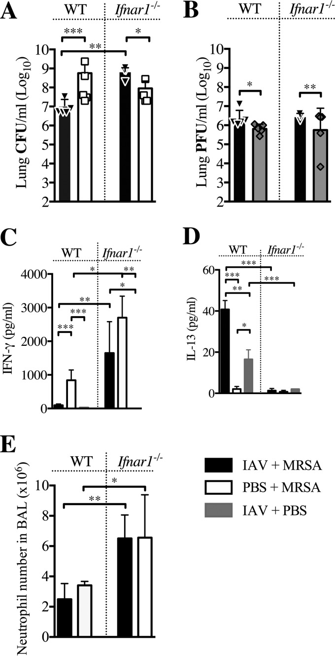
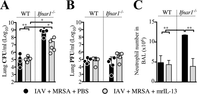
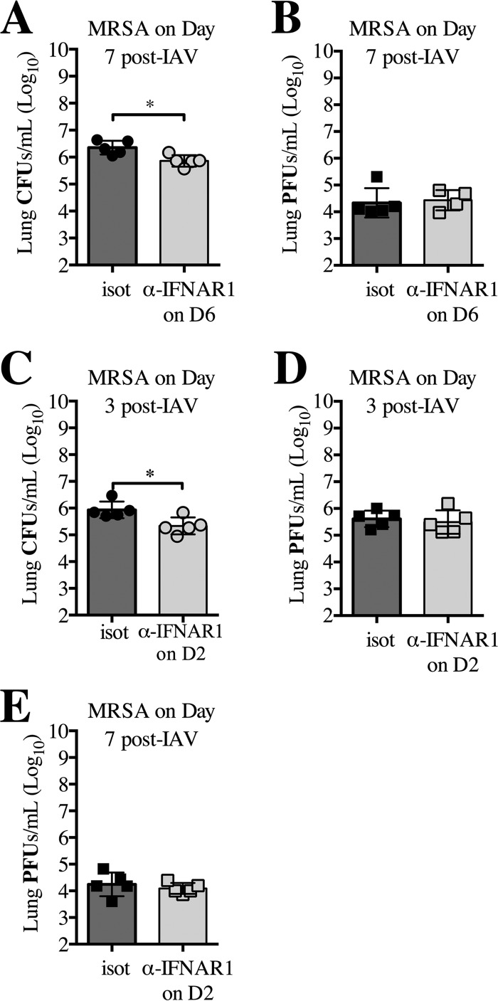
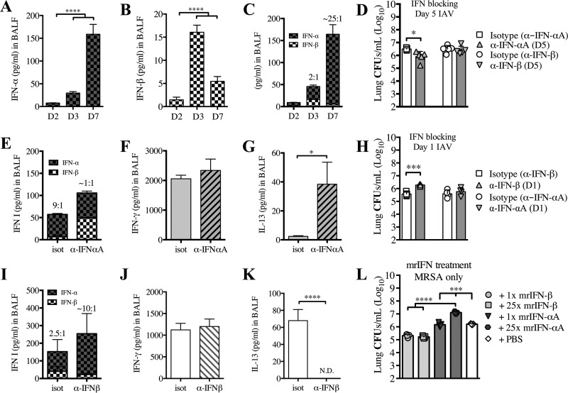
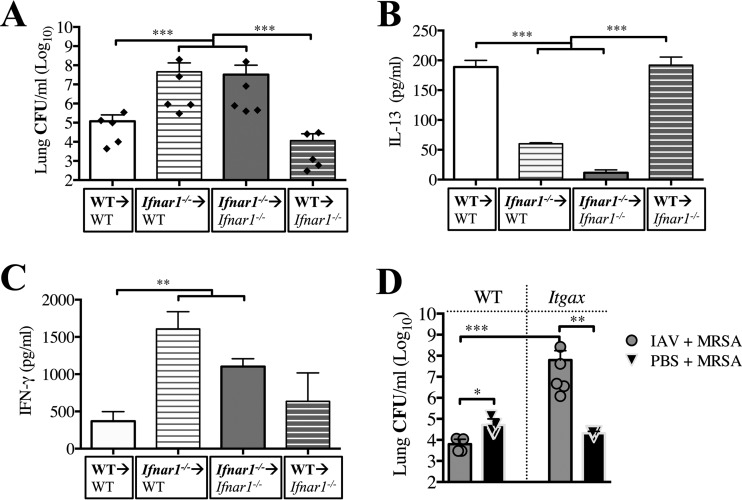
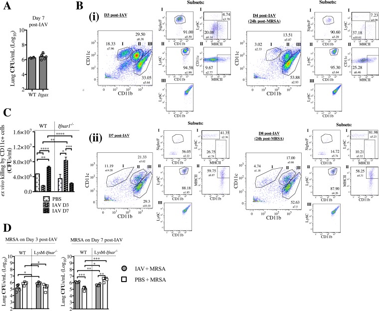
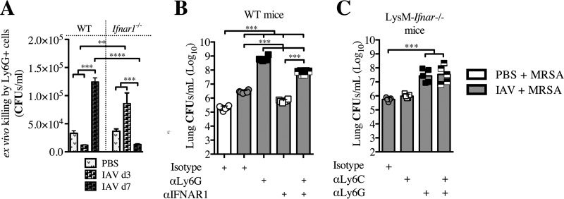
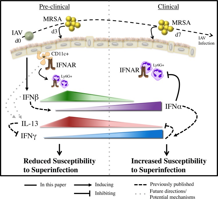
Bacterial superinfections are a primary cause of death during influenza pandemics and epidemics. Type I interferon (IFN) signaling contributes to increased susceptibility of mice to bacterial superinfection around day 7 post-influenza A virus (IAV) infection. Here we demonstrate that the reduced susceptibility to methicillin-resistant Staphylococcus aureus (MRSA) at day 3 post-IAV infection, which we previously reported was due to interleukin-13 (IL-13)/IFN-γ responses, is also dependent on type I IFN signaling and its subsequent requirement for protective IL-13 production. We found, through utilization of blocking antibodies, that reduced susceptibility to MRSA at day 3 post-IAV infection was IFN-β dependent, whereas the increased susceptibility at day 7 was IFN-α dependent. IFN-β signaling early in IAV infection was required for MRSA clearance, whereas IFN-α signaling late in infection was not, though it did mediate increased susceptibility to MRSA at that time. Type I IFN receptor (IFNAR) signaling in CD11c(+) and Ly6G(+) cells was required for the observed reduced susceptibility at day 3 post-IAV infection. Depletion of Ly6G(+) cells in mice in which IFNAR signaling was either blocked or deleted indicated that Ly6G(+) cells were responsible for the IFNAR signaling-dependent susceptibility to MRSA superinfection at day 7 post-IAV infection. Thus, during IAV infection, the temporal differences in type I IFN signaling increased bactericidal activity of both CD11c(+) and Ly6G(+) cells at day 3 and reduced effector function of Ly6G(+) cells at day 7. The temporal differential outcomes induced by IFN-β (day 3) and IFN-α (day 7) signaling through the same IFNAR resulted in differential susceptibility to MRSA at 3 and 7 days post-IAV infection.

Importance: Approximately 114,000 hospitalizations and 40,000 annual deaths in the United States are associated with influenza A virus (IAV) infections. Frequently, these deaths are due to community-acquired Gram-positive bacterial species, many of which show increasing resistance to antibiotic therapy. Severe complications, including parapneumonic empyema and necrotizing pneumonia, can arise, depending on virulence factors expressed by either the virus or bacteria. Unfortunately, we are unable to control the expression of these virulence factors, making host responses a logical target for therapeutic interventions. Moreover, interactions between virus, host, and bacteria that exacerbate IAV-related morbidities and mortalities are largely unknown. Here, we show that type I interferon (IFN) expression can modulate susceptibility to methicillin-resistant Staphylococcus aureus (MRSA) infection, with IFN-β reducing host susceptibility to MRSA infection while IFN-α increases susceptibility. Our data indicate that treatments designed to augment IFN-β and/or inhibit IFN-α production around day 7 post-IAV infection could reduce susceptibility to deadly superinfections.


Figures

FIG 1
FIG 1
Ifnar1 −/− mice are susceptible to secondary MRSA pneumonia early after IAV infection. Ifnar1−/− and C57BL/6 (WT) mice were infected with IAV on day 0 and MRSA on day 3. (A and B) Bacterial (A) and viral (B) loads were evaluated 24 h after MRSA challenge (day 4 post-IAV infection). (C and D) Levels of IFN-γ (C) and IL-13 (D) were evaluated in cell-free BALF collected at the time of sacrifice. (E) Numbers of neutrophils in the BALF were determined at the time of sacrifice via differential counts. ***, P < 0.001; **, P < 0.01; *, P < 0.05.
FIG 2
FIG 2
Lack of IL-13 production by superinfected Ifnar1−/− mice contributes to the increased susceptibility to superinfection early after IAV infection. WT and Ifnar1−/− mice were infected with IAV and treated either with 0.5 µg mrIL-13 or an equal volume of PBS at the time of challenge with MRSA on day 3 post-IAV infection, as well as 3 and 6 h later. Bacterial (A) and viral (B) loads as well as neutrophil numbers in the BALF (C) were evaluated 24 h after MRSA challenge (day 4 post-IAV infection). **, P < 0.01.
FIG 3
FIG 3
Inhibition of signaling through IFNAR1 at day 7 post-IAV infection reduces susceptibility to MRSA superinfection. WT mice were infected with IAV (day 0), treated with anti-IFNAR1 or isotype antibody on day 6 (D6), and challenged with MRSA on day 7. (A and B) Bacterial (A) and viral (B) loads were evaluated 24 h after MRSA challenge (day 8 post-IAV infection). (C to E) WT mice were infected with IAV on day 0, treated with anti-IFNAR1 antibody on day 2 (C, D, and E), and challenged with MRSA on day 3 (C and D). Bacterial (C) and viral (D) loads were evaluated 24 h after MRSA challenge (day 4 post-IAV infection). Viral load was measured in the lungs 7 days post-IAV infection (E). *, P < 0.05.
FIG 4
FIG 4
Reduction of IFN-β corresponds to increased susceptibility to post-IAV infection bacterial superinfection. WT mice were infected with IAV on day 0. Levels of IFN-α (A) and IFN-β (B) were evaluated in cell-free BALF collected at the time of sacrifice (days 2 [D2], 3, or 7 post-IAV challenge). (C) A combination of the results graphed in panels A and B, thus depicting ratios of IFN-α to IFN-β. (D to G) WT mice were infected with IAV on day 0, treated with anti-IFN-αA, anti-IFN-β, or isotype antibody on day 5, and challenged with MRSA on day 7. Bacterial burden (D) and the levels of IFN-α and IFN-β (E), IFN-γ (F), and IL-13 (G) were evaluated 24 h after MRSA challenge (day 8 post-IAV infection). (H to K) WT mice were infected with IAV on day 0, treated with anti-IFN-β, anti-IFN-αA, or isotype antibody on day 1, and challenged with MRSA on day 3. Bacterial burden (H) and the levels of IFN-α and IFN-β (I), IFN-γ (J), and IL-13 (K) were evaluated 24 h after MRSA challenge (day 4 post-IAV infection). (L) WT mice were treated with mrIFN-αA or mrIFN-β (1× dose was 104 IU; 25× dose was 2.5 × 10⁵ IU) 24 h prior to being infected with MRSA. The bacterial burden was evaluated 24 h after MRSA challenge. Data shown are mean ± SEM results for 4 mice from one experiment. ****, P < 0.0001, ***, P < 0.001; *, P < 0.05.
FIG 5
FIG 5
IFNAR signaling in BM-derived cells, specifically CD11c+ cells, is required for reduced susceptibility to superinfection early after IAV infection. BM chimeric mice were infected with IAV on day 0 and challenged with MRSA 3 days later (below each graph, the donor genotype is shown in bold, with the arrow indicating the recipient). (A) Lung bacterial burden was evaluated 24 h after MRSA challenge (day 4 post-IAV infection). (B and C) Levels of IL-13 (B) and IFN-γ (C) were evaluated in cell-free BALF collected at the time of sacrifice. (D) Itgax (Ifnar1fl/fl;Itgax-cre) and WT mice were infected with IAV (or inoculated with PBS) and challenged with MRSA (or PBS) 3 days later. Bacterial load was evaluated 24 h after MRSA challenge (day 4 post-IAV infection). ***, P < 0.001, **, P < 0.01, *, P < 0.05.
FIG 6
FIG 6
IFNAR signaling in CD11c+ cells is sufficient for killing MRSA at day 3 post-IAV infection, but not at day 7, and corresponds to phenotypic alterations occurring during IAV infection. (A) Itgax (Ifnar1fl/fl;Itgax-cre) mice were infected with IAV on day 0 and infected with MRSA on day 7. Bacterial burden was evaluated 24 h after MRSA challenge (day 8 post-IAV infection). (B) WT mice were sacrificed at the designated times post-IAV infection and post-MRSA infection (set i scatter plots, day 3 [D3] and day 4; set ii scatter plots, day 7 and day 8 post-IAV infection). Cells isolated from the BALF were stained and analyzed by using FACS. The gating for live cells was set on forward scatter versus side scatter. (i) In the left-most graph, staining for CD11c versus CD11b was determined by gating on the total live cells. The gating strategy entailed major gates (subsets) I (CD11c+ CD11b), II (CD11b+ CD11cint), and III (CD11b+ CD11c). The smaller groups of graphs for the subsets summarize scatter plots for SiglecF+, Ly6C+, and MHC-II+. Representative FACS plots with cell percentages (means and SEM) are shown. (C) WT and Ifnar1−/− mice were infected with IAV on day 0 and CD11c+ cells were purified from the lungs at days 3 and 7, and their ability to kill MRSA following a 1.5-h incubation ex vivo was determined. (D) WT and LysM-Ifnar (Ifnar1fl/fl;LysMcre/cre) mice were infected with IAV on day 0 and infected with MRSA on either day 3 or day 7. The bacterial burden was evaluated 24 h after MRSA challenge (day 4 or 8 post-IAV infection). ****, P < 0.0001; ***, P < 0.001; **, P < 0.01; *, P < 0.05.
FIG 7
FIG 7
IFNAR signaling impairs Ly6G+ cell ability to kill MRSA on day 7 post-IAV infection. (A) WT and Ifnar1−/− mice were infected with IAV on day 0, and Ly6G+ cells were purified from the lungs at day 3 and 7. The ability of these cells to kill MRSA following a 3-h incubation ex vivo was determined. (B) WT mice were infected with IAV on day 0 and treated with anti-IFNAR1 or isotype antibody on day 5.5 and/or anti-Ly6G or isotype antibody on day 6.5 and then infected with MRSA on day 7. Bacterial burden was evaluated 24 h after MRSA challenge (day 8 post-IAV infection). (C) LysM-Ifnar mice were infected with IAV on day 0, treated with antibody (anti-Ly6G, anti-Ly6C, or both) on day 6.5, and infected with MRSA on day 7. Bacterial burdens were evaluated 24 h after MRSA challenge (day 8 post-IAV infection). ***, P < 0.001.
FIG 8
FIG 8
A model representation of the results and potential mechanisms presented, demonstrating the unique and distinct type I IFN signaling responses involved in post-IAV infection MRSA superinfection. We specifically determined that susceptibility to superinfection corresponded to the relative ratio of IFN-α and IFN-β. We found that increased IFN-β during the preclinical stage of IAV infection (day 3 [d3]) correlated with reduced susceptibility to MRSA superinfection, whereas increased IFN-α during the clinical stage of IAV infection (day 7) correlated with increased susceptibility to MRSA superinfection. We determined that type I IFN signaling in CD11c+ cells was required for the reduced susceptibility to superinfection during the preclinical stage (day 3) of IAV infection, but not during the clinical stage (day 7). We also found that type I IFN signaling in Ly6G+ cells was required for killing MRSA in the preclinical stage; however, type I IFN signaling in Ly6G+ cells during the clinical stage was detrimental for the control and susceptibility to superinfection. Further, we determined that this type I IFN-dependent signaling is involved in the IL-13/IFN-γ response known to be involved in susceptibility to superinfection (8). Future research will involve determining how type I IFN signaling regulates the production of IL-13.

Similar articles

Cited by

References

    1. Metersky ML, Masterton RG, Lode H, File TM, Babinchak T. 2012. Epidemiology, microbiology, and treatment considerations for bacterial pneumonia complicating influenza. Int J Infect Dis 16:e321–e331. doi:10.1016/j.ijid.2012.01.003. - DOI - PubMed
    1. McCullers JA. 2014. The co-pathogenesis of influenza viruses with bacteria in the lung. Nat Rev Microbiol 12:252–262. doi:10.1038/nrmicro3231. - DOI - PubMed
    1. Chertow DS, Memoli MJ. 2013. Bacterial coinfection in influenza A grand rounds review. JAMA 309:275–282. doi:10.1001/jama.2012.194139. - DOI - PubMed
    1. Lee M-H, Arrecubieta C, Martin FJ, Prince A, Borczuk AC, Lowy FD. 2010. A postinfluenza model of Staphylococcus aureus pneumonia. J Infect Dis 201:508–515. doi:10.1086/650204. - DOI - PMC - PubMed
    1. Iverson AR, Boyd KL, McAuley JL, Plano LR, Hart ME, McCullers JA. 2011. Influenza virus primes mice for pneumonia from Staphylococcus aureus. J Infect Dis 203:880–888. doi:10.1093/infdis/jiq113. - DOI - PMC - PubMed

Publication types

MeSH terms