

Accelerated decline in white matter integrity in clinically normal individuals at risk for Alzheimer's disease
- PMID: 27143434
- PMCID: PMC4857135
- DOI: 10.1016/j.neurobiolaging.2016.03.016
Accelerated decline in white matter integrity in clinically normal individuals at risk for Alzheimer's disease
Abstract
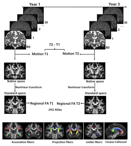
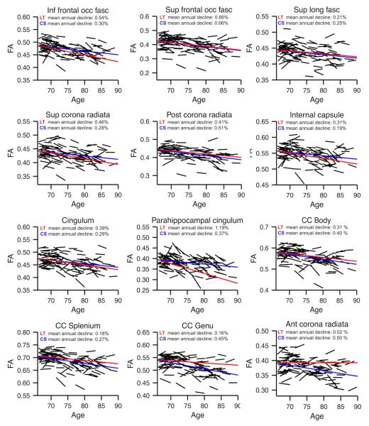
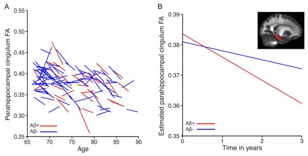
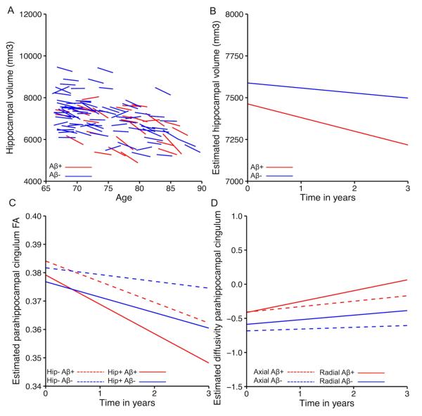
Prior studies have identified white matter abnormalities in Alzheimer's disease (AD). Yet, cross-sectional studies in normal older individuals show little evidence for an association between markers of AD risk (APOE4 genotype and amyloid deposition), and white matter integrity. Here, 108 normal older adults (age, 66-87) with assessments of apolipoprotein e4 (APOE4) genotype and assessment of amyloid burden by positron emission tomography underwent diffusion tensor imaging scans for measuring white matter integrity at 2 time points, on average 2.6 years apart. Linear mixed-effects models showed that amyloid burden at baseline was associated with steeper decline in fractional anisotropy in the parahippocampal cingulum (p < 0.05). This association was not significant between baseline measures suggesting that longitudinal analyses can provide novel insights that are not detectable in cross-sectional designs. Amyloid-related changes in hippocampus volume did not explain the association between amyloid burden and change in fractional anisotropy. The results suggest that accumulation of cortical amyloid and white matter changes in parahippocampal cingulum are not independent processes in individuals at increased risk for AD.
Keywords: Aging; Amyloid; Diffusion tensor imaging; Longitudinal; White matter.
Copyright © 2016 Elsevier Inc. All rights reserved.
Figures
References
-
- Barrick TR, Charlton RA, Clark CA, Markus HS. White matter structural decline in normal ageing: A prospective longitudinal study using tract-based spatial statistics. Neuroimage. 2010;51:565–77. - PubMed
-
- Bartzokis G, Sultzer D, Lu PH, Nuechterlein KH, Mintz J, Cummings JL. Heterogeneous age-related breakdown of white matter structural integrity: implications for cortical “disconnection” in aging and Alzheimer’s disease. Neurobiol Aging. 2004;25:843–51. - PubMed
-
- Bates D, Mächler M, Bolker B, Walker S. Fitting linear mixed-effects models using lme4. 2014. arXiv.
Publication types
MeSH terms
Substances
Grants and funding
- K24 AG035007/AG/NIA NIH HHS/United States
- P01 AG036694/AG/NIA NIH HHS/United States
- S10 RR019254/RR/NCRR NIH HHS/United States
- R01 AG027435/AG/NIA NIH HHS/United States
- P50 AG005134/AG/NIA NIH HHS/United States
- R01 AG034556/AG/NIA NIH HHS/United States
- HHMI/Howard Hughes Medical Institute/United States
- S10 RR019307/RR/NCRR NIH HHS/United States
- S10 RR023043/RR/NCRR NIH HHS/United States
- K01 AG040197/AG/NIA NIH HHS/United States
- P41 EB015896/EB/NIBIB NIH HHS/United States
- S10 RR023401/RR/NCRR NIH HHS/United States
LinkOut - more resources
Full Text Sources
Other Literature Sources
Medical
