. 2016 May 4:6:25252.
doi: 10.1038/srep25252.

Using Drosophila as an integrated model to study mild repetitive traumatic brain injury

Affiliations

Using Drosophila as an integrated model to study mild repetitive traumatic brain injury

Ayeh Barekat et al. Sci Rep. .

Abstract

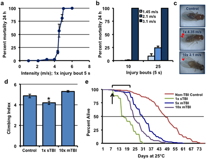
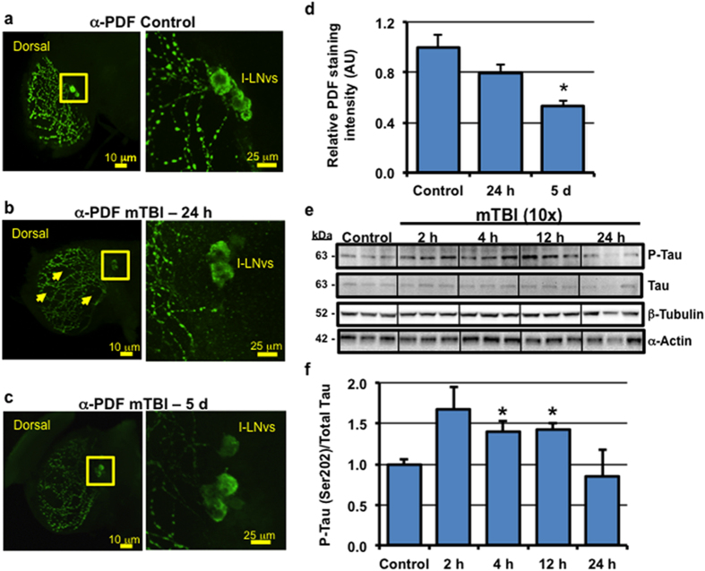
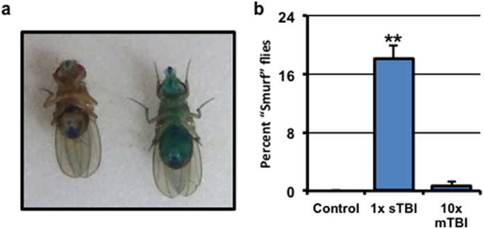
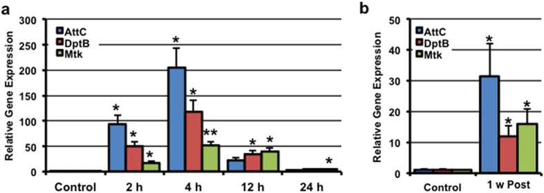
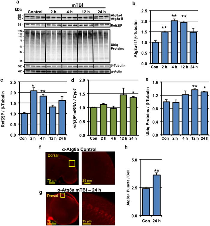
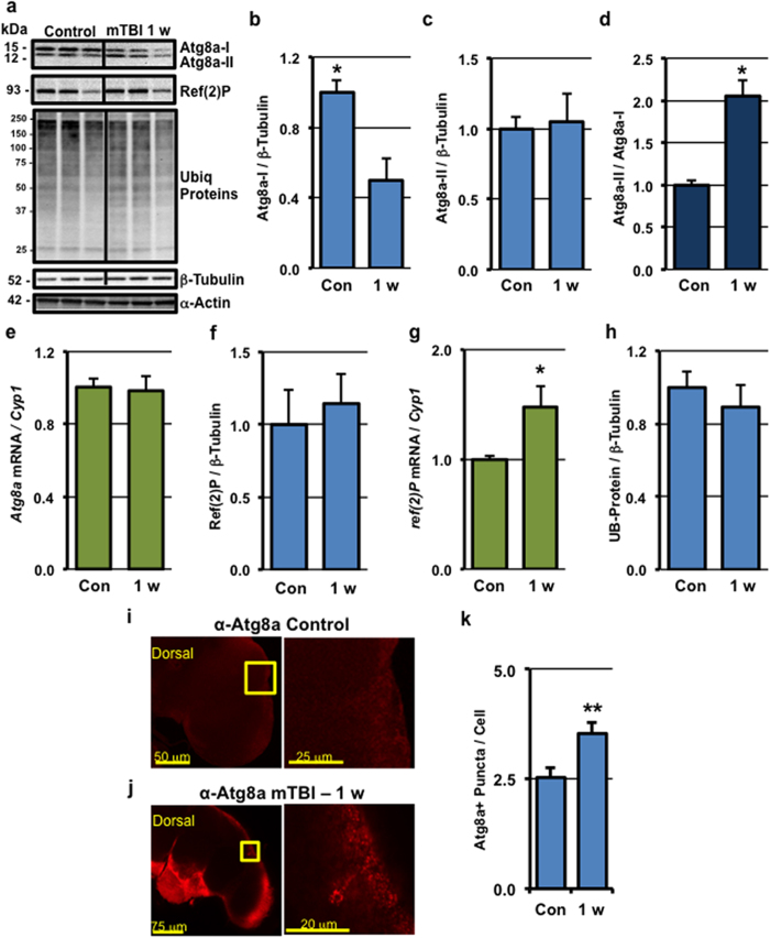
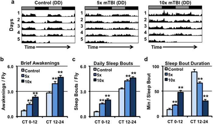
Traumatic brain injury (TBI) is a major cause of morbidity and mortality worldwide. In addition, there has been a growing appreciation that even repetitive, milder forms of TBI (mTBI) can have long-term deleterious consequences to neural tissues. Hampering our understanding of genetic and environmental factors that influence the cellular and molecular responses to injury has been the limited availability of effective genetic model systems that could be used to identify the key genes and pathways that modulate both the acute and long-term responses to TBI. Here we report the development of a severe and mild-repetitive TBI model using Drosophila. Using this system, key features that are typically found in mammalian TBI models were also identified in flies, including the activation of inflammatory and autophagy responses, increased Tau phosphorylation and neuronal defects that impair sleep-related behaviors. This novel injury paradigm demonstrates the utility of Drosophila as an effective tool to validate genetic and environmental factors that influence the whole animal response to trauma and to identify prospective therapies needed for the treatment of TBI.


Figures

Figure 1
Figure 1. Development of the mild TBI model in Drosophila.
(a) Male wild-type flies (1-week of age) were subjected to different injury intensities (5-second) using the Omni Bead Ruptor-24 and the number of dead flies (n ≥ 3; cohorts of 50 flies) was used to establish the 24 hour post-injury mortality. (b) Male flies (1-week of age) were subjected to multiple 5-second injury bouts at lower intensity settings and used to establish 24 hour post-injury mortality for multi-bout injuries (n ≥ 3; cohorts of 50 flies). (c) Representative images of flies following injury highlighting external damage to wing structures (red arrows). (d) The climbing index of groups of male wild-type control flies and flies subjected to a single sTBI (, 4.35 m/s intensity) or 10× mTBI (2.1 m/s intensity) injury bouts (1-week of age; cohorts of 25 flies; n ≥ 6). (e) Lifespan profiles of male wild-type control flies and flies subjected to (4.35 m/s intensity), or 10× (2.1 m/s intensity) injury bout(s) at 1-week of age (n ≥ 59, arrow indicates time of injury). The bracket highlights the 2-week delay in mortality in flies exposed to mTBI. See Materials and Methods for clarification of the injury protocol and Supplementary Table S1 for additional lifespan data and statistics. *P < 0.05.
Figure 2
Figure 2. Acute impact of mild TBI on neurons.
(a–c) The brains of adult male wild-type flies were dissected from (a) control flies (n = 15) and mTBI-treated (10×, 2.1 m/s) flies (b) after 24 hours (n = 11) and (c) 5 days (n = 9). Representative compressed confocal Z series and enlarged images (inset) of PDF staining patterns show the l-LNvs cell bodies and innervation patterns into the adult optic lobes. Arrows highlight damaged CNS regions that have lost PDF positive projections and synapses. (d) Quantification of PDF-staining intensity within the optic lobe (n ≥ 4 compressed Z-stacks of individual whole fly brains). (e) Replicate cohorts of male F1 flies with neuronal expression of the human Tau protein (APPL-Gal4/UAS-hTau) were exposed to 10× mTBI (2.1 m/s intensity) bouts at 1-week of age and collected at indicated time points. Western blots of phosphorylated-Tau (Ser 202), total Tau, α-Actin, and β-Tubulin from neural lysates of control and mTBI-treated flies. (f) Quantification of the results in (e), normalized to total Tau protein levels. *P < 0.05.
Figure 3
Figure 3. Intestinal integrity following TBI.
(a) Representative image of a female “Smurf” fly that displayed intestinal barrier dysfunction, as detected by blue dye permeation throughout the body (right). When the intestinal barrier was intact, the blue dye is mainly found in the proboscis and the digestive tract (left). (b) The percent of male wild-type flies with intestinal barrier dysfunction in groups of control flies and flies subjected to a single sTBI (, 4.35 m/s intensity) or 10× mTBI (2.1 m/s intensity) injury bouts (1-week of age; cohorts of 40 flies; n ≥ 6). **P < 0.01.
Figure 4
Figure 4. Innate immune response activation following mild TBI.
Replicate cohorts of wild-type male flies (1-week of age; cohorts of 25 flies; n ≥ 3) were exposed to 10× mTBI (2.1 m/s intensity) bouts. RNA was isolated from the heads of control and injured flies at the indicated time-points following mTBI during the (a) acute phase and at (b) 1 week post-injury (long-term). Expression of AttC, DptB, and Mtk were normalized using Cyp1 as the reference gene. *P < 0.05, **P < 0.01.
Figure 5
Figure 5. Acute impact of mild TBI on autophagic responses.
Replicate cohorts of wild-type male flies were exposed to 10× mTBI (2.1 m/s intensity) bouts at 1-week of age and collected at indicated time points. (a) Western blots of neural extracts were probed sequentially for the Atg8a, Ref(2)P, Ubiquitin (Ubiq) proteins, α-Actin, and β-Tubulin proteins. (b,c,e) Quantification of the results from (a) normalized to β-Tubulin levels. (d) ref(2)P mRNA expression of control and mTBI-treated flies at the indicated time points (cohorts of 25 flies; n = 3). (f,g) The brains from control (n = 15) and 10× mTBI-treated (2.1 m/s intensity) adult wild-type male flies (n = 10) were dissected at 24 hours post-injury. Representative and enlarged images (highlighted inset) of Atg8a staining patterns are shown in the optic lobe region of the adult CNS. (h) Atg8a-positive (Atg8a+) puncta were counted in neuronal cells found in the optic lobe region of the brain from images collected from (f,g). *P < 0.05, **P < 0.01.
Figure 6
Figure 6. Long-term impact of mild TBI on autophagic responses.
(a) Replicate cohorts of wild-type flies were exposed to 10× mTBI (2.1 m/s intensity) bouts at 1-week of age and collected 1-week post-injury (1 w). Western blots of neural extracts were probed for Atg8a, Ref(2)P, Ubiquitin (Ubiq) protiens, α-Actin, and β-Tubulin. (b,c,f,h) Quantification of the results from (a) normalized to β-Tubulin. (d) The ratio of Atg8a-II protein levels to Atg8a-I. (e,g) Atg8a and ref(2)P mRNA expression of control and mTBI-treated flies at the indicated time points, respectively (cohorts of 25 flies; n = 3). (I,j) The brains of adult male wild-type flies exposed to 10× mTBI (2.1 m/s intensity) bouts 1-week following injury (n = 12) and age-matched control flies (2-weeks of age, n = 15) were dissected. Representative and enlarged images (highlighted inset) of Atg8a staining patterns are shown. (k) Atg8a-positive (Atg8a+) puncta were counted in neuronal cells found in the optic lobe region of the brain from images collected from (I,j). *P < 0.05, **P < 0.01.
Figure 7
Figure 7. Long-term impact of mild TBI on Drosophila circadian and sleep behaviors.
Groups of wild-type female flies were exposed to or 10× mTBI (2.1 m/s intensity) bouts at 1-week of age and allowed to recover for 5-days before being placed into the DAM system and assessed in constant darkness (DD). (a) Representative double-plotted actograms of control (n = 58), 5× (n = 56), and 10× (n = 59) mTBI treated flies. Using the MATLAB-based software, analysis of sleep-related behaviors was performed to assess the number of (b) Brief Awakenings, (c) Daily Sleep Bouts, and (d) Sleep Bout Duration during the subjective day (CT0-12) and subjective night (CT12-24) time periods. **P < 0.01.

References

    1. Xiong Y., Mahmood A. & Chopp M. Animal models of traumatic brain injury. Nat Rev Neurosci 14, 128–142, doi: 10.1038/nrn3407 (2013). - DOI - PMC - PubMed
    1. Kane M. J. et al.. A mouse model of human repetitive mild traumatic brain injury. J Neurosci Methods 203, 41–49, doi: 10.1016/j.jneumeth.2011.09.003 (2012). - DOI - PMC - PubMed
    1. Mouzon B. et al.. Repetitive mild traumatic brain injury in a mouse model produces learning and memory deficits accompanied by histological changes. J Neurotrauma 29, 2761–2773, doi: 10.1089/neu.2012.2498 (2012). - DOI - PubMed
    1. Landre N., Poppe C. J., Davis N., Schmaus B. & Hobbs S. E. Cognitive functioning and postconcussive symptoms in trauma patients with and without mild TBI. Arch Clin Neuropsychol 21, 255–273, doi: 10.1016/j.acn.2005.12.007 (2006). - DOI - PubMed
    1. Stern R. A. et al.. Long-term consequences of repetitive brain trauma: chronic traumatic encephalopathy. PM R 3, S460–467, doi: 10.1016/j.pmrj.2011.08.008 (2011). - DOI - PubMed

Publication types

MeSH terms