. 2016 Jun 14;133(24):2447-58.
doi: 10.1161/CIRCULATIONAHA.116.021494. Epub 2016 Apr 25.

Prolyl-4 Hydroxylase 2 (PHD2) Deficiency in Endothelial Cells and Hematopoietic Cells Induces Obliterative Vascular Remodeling and Severe Pulmonary Arterial Hypertension in Mice and Humans Through Hypoxia-Inducible Factor-2α

Affiliations

Prolyl-4 Hydroxylase 2 (PHD2) Deficiency in Endothelial Cells and Hematopoietic Cells Induces Obliterative Vascular Remodeling and Severe Pulmonary Arterial Hypertension in Mice and Humans Through Hypoxia-Inducible Factor-2α

Zhiyu Dai et al. Circulation. .

Abstract

Background: Vascular occlusion and complex plexiform lesions are hallmarks of the pathology of severe pulmonary arterial hypertension (PAH) in patients. However, the mechanisms of obliterative vascular remodeling remain elusive; hence, current therapies have not targeted the fundamental disease-modifying mechanisms and result in only modest improvement in morbidity and mortality.

Methods and results: Mice with Tie2Cre-mediated disruption of Egln1 (encoding prolyl-4 hydroxylase 2 [PHD2]; Egln1(Tie2)) in endothelial cells and hematopoietic cells exhibited spontaneous severe PAH with extensive pulmonary vascular remodeling, including vascular occlusion and plexiform-like lesions, resembling the hallmarks of the pathology of clinical PAH. As seen in patients with idiopathic PAH, Egln1(Tie2) mice exhibited unprecedented right ventricular hypertrophy and failure and progressive mortality. Consistently, PHD2 expression was diminished in lung endothelial cells of obliterated pulmonary vessels in patients with idiopathic PAH. Genetic deletions of both Egln1 and Hif1a or Egln1 and Hif2a identified hypoxia-inducible factor-2α as the critical mediator of the severe PAH seen in Egln1(Tie2) mice. We also observed altered expression of many pulmonary hypertension-causing genes in Egln1(Tie2) lungs, which was normalized in Egln1(Tie2)/Hif2a(Tie2) lungs. PHD2-deficient endothelial cells promoted smooth muscle cell proliferation in part through hypoxia-inducible factor-2α-activated CXCL12 expression. Genetic deletion of Cxcl12 attenuated PAH in Egln1(Tie2) mice.

Conclusions: These studies defined an unexpected role of PHD2 deficiency in the mechanisms of severe PAH and identified the first genetically modified mouse model with obliterative vascular remodeling and pathophysiology recapitulating clinical PAH. Thus, targeting PHD2/hypoxia-inducible factor-2α signaling is a promising strategy to reverse vascular remodeling for treatment of severe PAH.

Keywords: endothelial cells; plexiform lesion; pulmonary heart disease; pulmonary hypertension; right heart hypertrophy and failure; vascular remodeling; vascular smooth muscle cells.


Figures

Figure 1
Figure 1
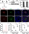
Spontaneous severe PAH in Egln1Tie2 mice. (A–D) Tie2Cre-mediated disruption of Egln1 in lung ECs. A diagram showing the strategy for generation of Egln1Tie2 mice (A). Representative Western blotting demonstrating diminished PHD2 protein expression in EC lysate isolated from Egln1Tie2 lungs compared to WT (B). The experiment was repeated twice with similar data. QRT-PCR analysis demonstrating PHD2 deletion in Egln1Tie2 lung ECs but not in fibroblasts (Fib) (C). Data are expressed mean ±SD (n=3) (C). Representative micrographs of immunostaining showing EC-specific disruption of PHD2 in Egln1Tie2 mouse lungs. Lung tissues were co-stained with anti-PHD2 and anti-CD31 (marker for ECs). Nuclei were counterstained with DAPI. PHD2 expression was diminished in pulmonary vascular ECs but not bronchial epithelial cells in Egln1Tie2 lungs. Please note the marked vascular remodeling in Egln1Tie2 lungs. Br, bronchiole; V, vessel. Scale bar, 50 µm (D). CKO = Egln1Tie2. (E) Representative RVSP tracings (each=1 s). (F) Dramatic increase of RVSP in Egln1Tie2 mice. Bars represent the mean. (G) Marked RV hypertrophy in Egln1Tie2 mice. **, P < 0.01; ***, P < 0.001 (Student’s t test: C; Two-way ANOVA followed by Games-Howell post hoc analysis: F and G).
Figure 2
Figure 2
Severe RV hypertrophy and failure and progressive mortality of Egln1Tie2 mice. (A) Representative echocardiography showing an enlarged RV chamber and thickened RV wall (hypertrophy) in Egln1Tie2 mice (3.5 mo old). (B–E) Echocardiography demonstrating enlarged RV chamber at end systole (ES) and end diastole (ED) (B); increased RV wall thickness diastole (RVWTD) (C); decreased RV fraction area change (RVFAC), indicative of decreased RV contractility (D); and a decreased PA AT/ET ratio (E) in Egln1Tie2 mice (3.5 months old). Data are expressed as mean ± SD (n=5 WT and 6 Egln1Tie2). (F) QRT-PCR analysis demonstrating reactivation of an embryonic gene program in the right ventricles of Egln1Tie2 mice aged 3.5 mo, indicative of heart failure. ANF=Atrial natriuretic factor; sk-actin=skeletal α-actin. (G). Progressive mortality of Egln1Tie2 mice. *, P < 0.05; **, P < 0.01; ***, P < 0.001. Student’s t test (B-E), Welch t test (F) and log-rank (Mantel-Cox) test (G) were used for statistical analysis.
Figure 3
Figure 3
Attenuated PAH in Egln1Tie2 chimeric mice reconstituted with WT bone marrow cells. (A) A cartoon showing bone marrow (WT or CKO BM) transplantation to lethally irradiated WT mice. (B) QPCR analysis of Sry sequence (Y chromosome-specific gene) demonstrating greater than 95% efficiency of bone marrow reconstitution of female recipient WT mice with male Egln1Tie2 bone marrow. Bone marrow DNA from male WT mice was used as positive control. (C, D) Reconstitution of lethally irradiated WT mice with bone marrow cells from Egln1Tie2 mice (CKO BM) had no effects on RVSP (C) and RV/LV+S ratio (D). As controls, WT bone marrow cells were also transplanted to WT recipient mice. (E-G) Egln1Tie2 chimeric mice reconstituted with WT bone marrow cells exhibited marked decreases of RVSP and RV hypertrophy. ***, P < 0.001 (Student’s t test). CKO=Egln1Tie2.
Figure 4
Figure 4
Occlusive pulmonary vascular remodeling in Egln1Tie2 mice. (A) Representative micrographs of Russel-Movat pentachrome staining demonstrating thickening of the intima, medial, and adventitial, occlusion of the large and small vessels (black arrowheads), and plexiform-like lesions (red arrowheads) in 3.5 mo old Egln1Tie2 mice (n=3 WT and 6 Egln1Tie2 mice). Br, bronchus; V, vessel. Scale bar: 50 µm. (B) Anti-CD31 immunohistochemistry showing multiple-channel lesions positive for the endothelial marker CD31 (arrows). Scale bar: 50 µm. (C) Quantification of pulmonary vascular remodeling in Egln1Tie2 mice. Grade 1 (G1), medial hypertrophy; G2, medial hypertrophy and intimal thickening (partial occlusion); G3, occlusive lesions (>75% occlusion); G4, plexiform-like lesions. Much severe pulmonary vascular remodeling was identified in Egln1Tie2 mice at age of 3.5 mo compared to 1.5 mo. Total= all vessels quantified (n=280 vessels/group, n=5 mice/group). In 3.5 mo old Egln1Tie2 mice, plexiform-like lesions (G4) were mainly seen in large vessels (>100 µm diameter) whereas occlusive lesions (G3) were prominent in small vessels. (D-F) Representative micrographs showing anti-smooth muscle α-actin (α-SMA), anti-FSP1 or CD11b staining in pulmonary vascular lesions of Egln1Tie2 mice. . Br, bronchus; V, vessel. Scale bar, 50µm. (G) Representative micrographs showing proliferating ECs, SMCs and adventitial cells in pulmonary vascular lesions of Egln1Tie2 mice (3.5 mo old). Lung sections were immunostained with anti-Ki67 for cell proliferation and anti-CD31 for ECs. Arrowheads point to proliferating ECs; arrows indicate proliferating SMCs; open arrows denote proliferating adventitial cells. Scale bar, 50 µm. (H) Western blotting demonstrating increased expression of PCNA, a marker for cell proliferation in Egln1Tie2 lungs.
Figure 5
Figure 5
Diminished PHD2 expression in occlusive pulmonary vessels of IPAH patients. (A) Immunostaining demonstrating diminished PHD2 expression (red) in the lumen of occlusive vessels (arrowheads) of IPAH lungs. Lung sections exhibited strong autofluorescence (AutoF) which helped to show the morphology. Arrows point to non-occlusive vessels; asterisk indicates blood cells. Scale bar, 50 µm. (B) Quantification of PHD2 expression. Immunofluorescent intensity (Fluo) was graded from 1 to 10 with 10 the highest. PHD2 expression was diminished in occlusive vessels of IPAH patients. Data are expressed as mean ± SD. **, P < 0.01 (Welch t test). A.U., arbitrary units.
Figure 6
Figure 6
Role of HIF-2α activation in Egln1Tie2 mice in mediating severe PAH. (A) Western blot showing stabilized HIF-1α and HIF-2α expression in Egln1Tie2 mouse lungs. The experiment was repeated twice with similar data. (B-D) Genetic deletion of HIF2α but not HIF1α in Egln1Tie2 mice completely normalized RVSP (C) and inhibited RV hypertrophy evident by normalized RV/LV+S ratio (2 months old). (D) Bars represent the mean. (E, F) Echocardiography demonstrating normalization of the RVWTD (E) and the PA AT/ET ratio (F) in EH2 mice at age of 3.5 months. Data are expressed as mean ± SD (n=6 WT, 8 Egln1Tie2 and 5 EH2). (G) Russel-Movat pentachrome staining showed normal pulmonary vascular structure in EH2 mice at 3.5 month of age in contrast to Egln1Tie2 mice. Br, bronchiole; V, vessel. Scale bar: 50 µm. (H) Representative micrographs showing increased muscularization of distal pulmonary vessels (<50µm in diameter) in Egln1Tie2 mice and normalization in EH2 mice aged 3.5 months. Lung sections were immunostained with anti-smooth muscle α-actin (SMA) (red). Arrows point to muscularized distal vessels. Scale bar, 50 µm. (I) Quantification of muscularization. SMA– positive vessels were counted in 40 fields (×200) from each lung section. Data are expressed as mean ± SD (n=5 mice/group). *, P < 0.05; ***, P < 0.001. ANOVA followed by Games-Howell post hoc analysis was used for statistical analysis.
Figure 7
Figure 7
Expression profiling of genes associated with various PH animal models. (A) Representative heat map of RNA-seq analysis in WT, Egln1Tie2 (CKO) and EH2 mouse lungs (n=3 mice/group). (B-D) RNA-seq analysis of HIF-α target genes (B), PH-causing genes (C, D) in mouse lungs. (E) Representative diagram showing increased Cxcl12 expression in Egln1Tie2 lungs whereas inhibited in EH2 lungs identified by RNA-seq analysis. (F) QRT-PCR analysis of expression of genes known upregulated in published PH animal models. Data are expressed as mean ± SD (n=4 WT, 6 CKO and 6 EH2). (G) QRT-PCR analysis of expression of genes known downregulated in PH animal models. *, P < 0.05; **, P < 0.01; ***, P < 0.001. ANOVA followed by Games-Howell post hoc analysis was used for statistical analysis.
Figure 8
Figure 8
HIF-2α-mediated induction of CXCL12 in lung ECs induced SMC proliferation and contributed to severe PAH in Egln1Tie2 mice. (A) QRT-PCR analysis demonstrating HIF- 2α-dependent induction of CXCL12 in PHD2-deficient ECs. Small interfering RNAs specific for either PHD2, HIF1A, or HIF2A were transfected to human lung microvascular ECs. Scramble RNA against PHD2 was also transfected as a control. Data are expressed as mean ± SD (n=3). (B) Representative micrographs showing PHD2-deficient ECs induced SMC proliferation employing the Transwell co-culture system. Proliferating SMCs were immunostained with anti-BrdU antibody (green). Scale bar, 50 µm. (C) Quantification showing PHD2-deficient ECs induced a marked increase of SMC proliferation which was partially inhibited by knockdown of CXCL12. n=3. (D) QRT-PCR analysis demonstrating siRNA- mediated knockdown of CXCL12 in human lung ECs. (E) Cxcl12 expression was markedly induced in Egln1Tie2 (CKO) lungs in a HIF-2α but not HIF-1α-dependent manner. Lung tissues were collected from 1.5 mo old mice. (F) Generation of a mouse model with genetic deletions of both Egln1 and Cxcl12 (ECx). (G) ECx mice exhibited markedly decreased RVSP compared to CKO mice (3.5 mo old). (H) RV hypertrophy was also partially inhibited in ECx mice. **, P < 0.01; ***, P < 0.001. ANOVA followed by Games-Howell post hoc analysis was used for statistical analysis (A, C, E). Student’s t test was used for statistical analysis (D, G, H). (I) A diagram showing the model that PHD2 deficiency in ECs and HCs induces obliterative pulmonary vascular remodeling and severe PAH. Endothelial PHD2 deficiency is essential for the development of PAH while PHD2 deficiency in HCs promotes the severity of the disease. Through HIF-2α activation, PHD2 deficiency induces dysregulation of multiple signaling pathways in mouse lungs which may act together to induce severe pulmonary vascular remodeling including vascular occlusion and formation of plexiform-like lesions as well as vasoconstriction and thereby severe PAH and RV failure.

References

    1. McLaughlin VV, Archer SL, Badesch DB, Barst RJ, Farber HW, Lindner JR, Mathier MA, McGoon MD, Park MH, Rosenson RS, Rubin LJ, Tapson VF, Varga J, Harrington RA, Anderson JL, Bates ER, Bridges CR, Eisenberg MJ, Ferrari VA, Grines CL, Hlatky MA, Jacobs AK, Kaul S, Lichtenberg RC, Lindner JR, Moliterno DJ, Mukherjee D, Pohost GM, Rosenson RS, Schofield RS, Shubrooks SJ, Stein JH, Tracy CM, Weitz HH, Wesley DJ ACCF/AHA. ACCF/AHA 2009 expert consensus document on pulmonary hypertension: a report of the American College of Cardiology Foundation Task Force on Expert Consensus Documents and the American Heart Association: developed in collaboration with the American College of Chest Physicians, American Thoracic Society, Inc., and the Pulmonary Hypertension Association. Circulation. 2009;119:2250–2294. - PubMed
    1. Voelkel NF, Cool C. Pathology of pulmonary hypertension. Cardiol Clin. 2004;22:343–351. v. - PubMed
    1. Pietra GG, Capron F, Stewart S, Leone O, Humbert M, Robbins IM, Reid LM, Tuder RM. Pathologic assessment of vasculopathies in pulmonary hypertension. J Am Coll Cardiol. 2004;43:25S–32S. - PubMed
    1. Meyrick B. The pathology of pulmonary artery hypertension. Clin Chest Med. 2001;22:393–404. vii. - PubMed
    1. Stacher E, Graham BB, Hunt JM, Gandjeva A, Groshong SD, McLaughlin VV, Jessup M, Grizzle WE, Aldred MA, Cool CD, Tuder RM. Modern age pathology of pulmonary arterial hypertension. Am J Respir Crit Care Med. 2012;186:261–272. - PMC - PubMed

Publication types

MeSH terms

Substances