

Role of the mitochondrial DNA replication machinery in mitochondrial DNA mutagenesis, aging and age-related diseases
- PMID: 27143693
- PMCID: PMC5086445
- DOI: 10.1016/j.arr.2016.04.006
Role of the mitochondrial DNA replication machinery in mitochondrial DNA mutagenesis, aging and age-related diseases
Abstract
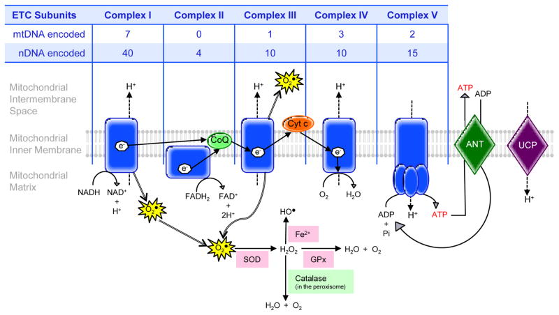
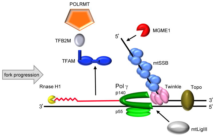
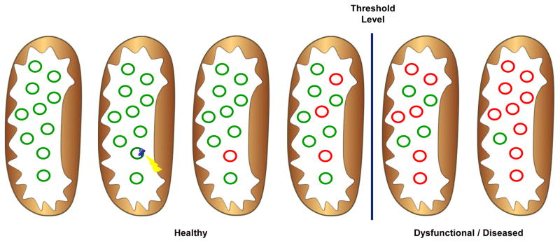
As regulators of bioenergetics in the cell and the primary source of endogenous reactive oxygen species (ROS), dysfunctional mitochondria have been implicated for decades in the process of aging and age-related diseases. Mitochondrial DNA (mtDNA) is replicated and repaired by nuclear-encoded mtDNA polymerase γ (Pol γ) and several other associated proteins, which compose the mtDNA replication machinery. Here, we review evidence that errors caused by this replication machinery and failure to repair these mtDNA errors results in mtDNA mutations. Clonal expansion of mtDNA mutations results in mitochondrial dysfunction, such as decreased electron transport chain (ETC) enzyme activity and impaired cellular respiration. We address the literature that mitochondrial dysfunction, in conjunction with altered mitochondrial dynamics, is a major driving force behind aging and age-related diseases. Additionally, interventions to improve mitochondrial function and attenuate the symptoms of aging are examined.
Keywords: Age-related diseases; Aging; DNA polymerase gamma; Mitochondrial DNA mutations; MtDNA replication; POLG.
Published by Elsevier B.V.
Figures

Similar articles
-
Mitochondrial function and mitochondrial DNA maintenance with advancing age.Biogerontology. 2014;15(5):417-38. doi: 10.1007/s10522-014-9515-2. Epub 2014 Jul 12. Biogerontology. 2014. PMID: 25015781 Review.
-
Ultra-sensitive sequencing reveals an age-related increase in somatic mitochondrial mutations that are inconsistent with oxidative damage.PLoS Genet. 2013;9(9):e1003794. doi: 10.1371/journal.pgen.1003794. Epub 2013 Sep 26. PLoS Genet. 2013. PMID: 24086148 Free PMC article.
-
Somatic mitochondrial DNA mutations in mammalian aging.Annu Rev Biochem. 2010;79:683-706. doi: 10.1146/annurev-biochem-060408-093701. Annu Rev Biochem. 2010. PMID: 20350166 Review.
-
The mtDNA mutation spectrum in the PolG mutator mouse reveals germline and somatic selection.BMC Genom Data. 2021 Nov 26;22(1):52. doi: 10.1186/s12863-021-01005-x. BMC Genom Data. 2021. PMID: 34823474 Free PMC article.
-
The mitochondrial DNA polymerase in health and disease.Subcell Biochem. 2010;50:211-22. doi: 10.1007/978-90-481-3471-7_11. Subcell Biochem. 2010. PMID: 20012584 Free PMC article. Review.
Cited by
-
Dynamic features of human mitochondrial DNA maintenance and transcription.Front Cell Dev Biol. 2022 Sep 7;10:984245. doi: 10.3389/fcell.2022.984245. eCollection 2022. Front Cell Dev Biol. 2022. PMID: 36158192 Free PMC article. Review.
-
The Signaling of Cellular Senescence in Diabetic Nephropathy.Oxid Med Cell Longev. 2019 Oct 3;2019:7495629. doi: 10.1155/2019/7495629. eCollection 2019. Oxid Med Cell Longev. 2019. PMID: 31687085 Free PMC article. Review.
-
Is There Still Any Role for Oxidative Stress in Mitochondrial DNA-Dependent Aging?Genes (Basel). 2018 Mar 21;9(4):175. doi: 10.3390/genes9040175. Genes (Basel). 2018. PMID: 29561808 Free PMC article. Review.
-
GDF15 and Cardiac Cells: Current Concepts and New Insights.Int J Mol Sci. 2021 Aug 18;22(16):8889. doi: 10.3390/ijms22168889. Int J Mol Sci. 2021. PMID: 34445593 Free PMC article. Review.
-
Single-molecule imaging of genome maintenance proteins encountering specific DNA sequences and structures.DNA Repair (Amst). 2023 Aug;128:103528. doi: 10.1016/j.dnarep.2023.103528. Epub 2023 Jun 24. DNA Repair (Amst). 2023. PMID: 37392578 Free PMC article. Review.
References
-
- Ahlqvist KJ, Hamalainen RH, Yatsuga S, Uutela M, Terzioglu M, Gotz A, Forsstrom S, Salven P, Angers-Loustau A, Kopra OH, Tyynismaa H, Larsson NG, Wartiovaara K, Prolla T, Trifunovic A, Suomalainen A. Somatic progenitor cell vulnerability to mitochondrial DNA mutagenesis underlies progeroid phenotypes in Polg mutator mice. Cell Metab. 2012;15:100–109. - PubMed
-
- Alvarez V, Corao AI, Sanchez-Ferrero E, De Mena L, Alonso-Montes C, Huerta C, Blazquez M, Ribacoba R, Guisasola LM, Salvador C, Garcia-Castro M, Coto E. Mitochondrial transcription factor A (TFAM) gene variation in Parkinson’s disease. Neurosci Lett. 2008;432:79–82. - PubMed
-
- Baris OR, Ederer S, Neuhaus JF, von Kleist-Retzow JC, Wunderlich CM, Pal M, Wunderlich FT, Peeva V, Zsurka G, Kunz WS, Hickethier T, Bunck AC, Stockigt F, Schrickel JW, Wiesner RJ. Mosaic Deficiency in Mitochondrial Oxidative Metabolism Promotes Cardiac Arrhythmia during Aging. Cell Metab. 2015;21:667–677. - PubMed
Publication types
MeSH terms
Substances
Grants and funding
LinkOut - more resources
Full Text Sources
Other Literature Sources
Medical
Research Materials
