Pirfenidone inhibits fibrosis in foreign body reaction after glaucoma drainage device implantation
- PMID: 27143855
- PMCID: PMC4841429
- DOI: 10.2147/DDDT.S99957
Pirfenidone inhibits fibrosis in foreign body reaction after glaucoma drainage device implantation
Abstract
Background: The aim of this study was to investigate the antiscarring effects of pirfenidone on foreign body reaction in a rabbit model of glaucoma drainage implant surgery.
Methods: Adult New Zealand White rabbits had glaucoma drainage device implantation using Model FP8 Ahmed glaucoma valves. One eye was randomly assigned to receive postoperative intrableb injection of pirfenidone followed by topical treatment. The other eye underwent the same procedure but without the addition of pirfenidone. Histochemical staining and immunohistochemistry for blebs were performed.
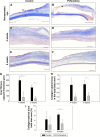
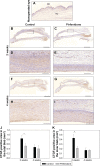
Results: The degree of cellularity was smaller in the pirfenidone group than in the control group at 2 weeks post operation (P=0.005). A few foreign body giant cells were detected in the inner border of the capsule, and their numbers were similar in the control and pirfenidone groups (P>0.05). Using Masson's trichrome stain, the inner collagen-rich layer was found to be thinner in the pirfenidone group than the control group at 4 weeks (P=0.031) and 8 weeks (P=0.022) post operation. The percentage of proliferating cell nuclear antigen-positive cells was lower in the pirfenidone group than in the control group at 2 weeks post operation (total bleb, P=0.022; inner bleb, P=0.036). Pirfenidone treatment decreased the immunoreactivity of connective tissue growth factor at 2 weeks post operation (total bleb, P=0.029; inner bleb, P=0.018). The height and area of α-smooth muscle actin expression were lower in the pirfenidone group than the control group at 2 weeks, 4 weeks, and 8 weeks post operation (all P<0.05).
Conclusion: Postoperative intrableb injection of pirfenidone followed by topical administration reduced fibrosis following glaucoma drainage device implantation. These findings suggest that pirfenidone may function as an antiscarring treatment in foreign body reaction after tube-shunt surgery.
Keywords: bleb; fibrosis; glaucoma drainage device surgery; transforming growth factor-β.
Figures


Similar articles
-
Foreign body reaction in glaucoma drainage implant surgery.Invest Ophthalmol Vis Sci. 2013 Jun 6;54(6):3957-64. doi: 10.1167/iovs.12-11310. Invest Ophthalmol Vis Sci. 2013. PMID: 23674756
-
Evaluation of pirfenidone as a new postoperative antiscarring agent in experimental glaucoma surgery.Invest Ophthalmol Vis Sci. 2011 May 16;52(6):3136-42. doi: 10.1167/iovs.10-6240. Invest Ophthalmol Vis Sci. 2011. PMID: 21330661
-
Application of 5-Fluorouracil-Polycaprolactone Sustained-Release Film in Ahmed Glaucoma Valve Implantation Inhibits Postoperative Bleb Scarring in Rabbit Eyes.PLoS One. 2015 Nov 18;10(11):e0141467. doi: 10.1371/journal.pone.0141467. eCollection 2015. PLoS One. 2015. PMID: 26579716 Free PMC article.
-
Rho kinase inhibitor AMA0526 improves surgical outcome in a rabbit model of glaucoma filtration surgery.Prog Brain Res. 2015;220:283-97. doi: 10.1016/bs.pbr.2015.04.014. Epub 2015 Jun 30. Prog Brain Res. 2015. PMID: 26497796 Review.
-
Pirfenidone: A Promising Drug in Ocular Therapeutics.Chem Biodivers. 2024 Mar;21(3):e202301389. doi: 10.1002/cbdv.202301389. Epub 2024 Feb 23. Chem Biodivers. 2024. PMID: 38299764 Review.
Cited by
-
Nanofiber-based glaucoma drainage implant improves surgical outcomes by modulating fibroblast behavior.Bioeng Transl Med. 2023 Jan 18;8(3):e10487. doi: 10.1002/btm2.10487. eCollection 2023 May. Bioeng Transl Med. 2023. PMID: 37206200 Free PMC article.
-
Local Delivery of Pirfenidone by PLA Implants Modifies Foreign Body Reaction and Prevents Fibrosis.Biomedicines. 2021 Jul 21;9(8):853. doi: 10.3390/biomedicines9080853. Biomedicines. 2021. PMID: 34440057 Free PMC article.
-
Development of a biodegradable antifibrotic local drug delivery system for glaucoma microstents.Biosci Rep. 2018 Aug 31;38(4):BSR20180628. doi: 10.1042/BSR20180628. Print 2018 Aug 31. Biosci Rep. 2018. PMID: 30061178 Free PMC article.
-
[Pirfenidone alleviates urethral stricture following urethral injury in rats by suppressing TGF-β1 signaling and inflammatory response].Nan Fang Yi Ke Da Xue Xue Bao. 2022 Mar 20;42(3):411-417. doi: 10.12122/j.issn.1673-4254.2022.03.14. Nan Fang Yi Ke Da Xue Xue Bao. 2022. PMID: 35426806 Free PMC article. Chinese.
-
Fighting Bleb Fibrosis After Glaucoma Surgery: Updated Focus on Key Players and Novel Targets for Therapy.Int J Mol Sci. 2025 Mar 5;26(5):2327. doi: 10.3390/ijms26052327. Int J Mol Sci. 2025. PMID: 40076946 Free PMC article.
References
-
- Almasieh M, Wilson AM, Morquette B, Cueva Vargas JL, Di Polo A. The molecular basis of retinal ganglion cell death in glaucoma. Prog Retin Eye Res. 2012;31(2):152–181. - PubMed
-
- Chen PP, Yamamoto T, Sawada A, Parrish RK, 2nd, Kitazawa Y. Use of antifibrosis agents and glaucoma drainage devices in the American and Japanese Glaucoma Societies. J Glaucoma. 1997;6(3):192–196. - PubMed
-
- Joshi AB, Parrish RK, 2nd, Feuer WF. 2002 survey of the American Glaucoma Society: practice preferences for glaucoma surgery and antifibrotic use. J Glaucoma. 2005;14(2):172–174. - PubMed
-
- Desai MA, Gedde SJ, Feuer WJ, Shi W, Chen PP, Parrish RK., 2nd Practice preferences for glaucoma surgery: a survey of the American Glaucoma Society in 2008. Ophthalmic Surg Lasers Imaging. 2011;42(3):202–208. - PubMed
Publication types
MeSH terms
Substances
LinkOut - more resources
Full Text Sources
Other Literature Sources

