. 2016 Apr 18:11:1577-91.
doi: 10.2147/IJN.S90850. eCollection 2016.

Potential antitumor activity of novel DODAC/PHO-S liposomes

Affiliations

Potential antitumor activity of novel DODAC/PHO-S liposomes

Arthur Cássio de Lima Luna et al. Int J Nanomedicine. .

Abstract

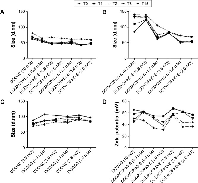
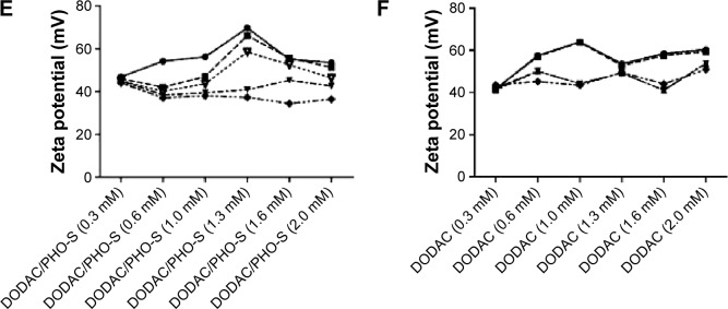
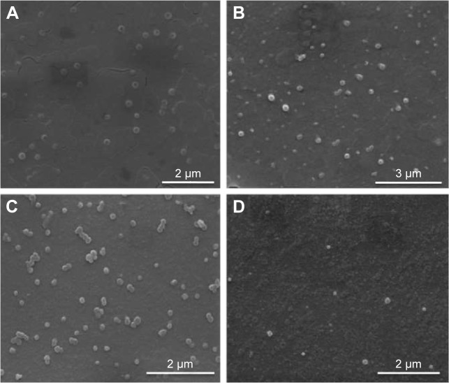
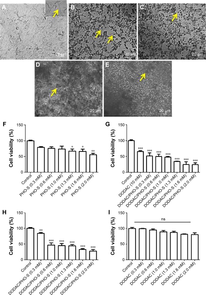
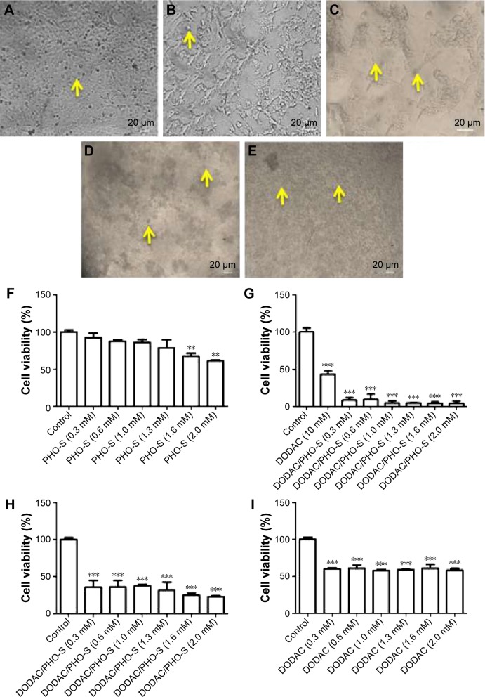
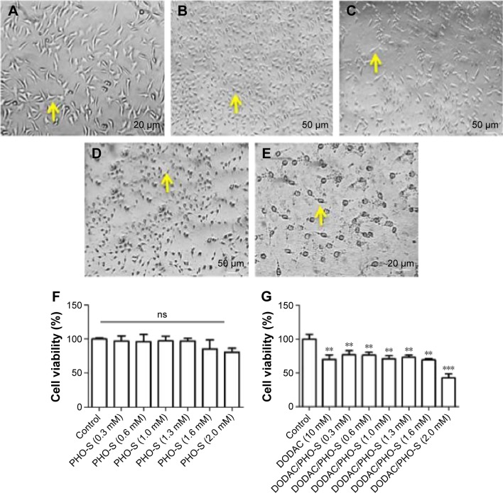
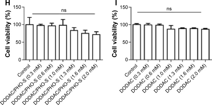
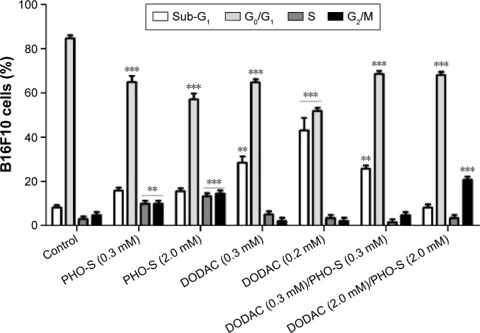
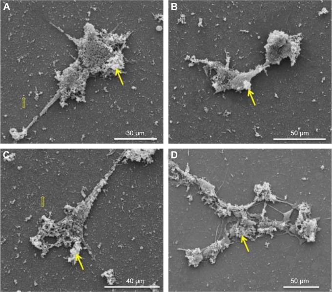
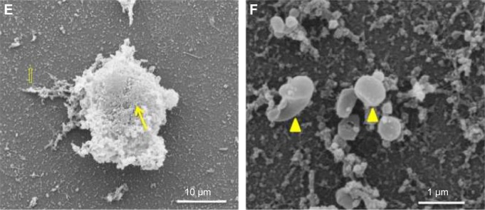
In recent studies, we showed that synthetic phosphoethanolamine (PHO-S) has a great potential for inducing cell death in several tumor cell lines without damage to normal cells. However, its cytotoxic effect and selectivity against tumor cells could increase with encapsulation in cationic liposomes, such as dioctadecyldimethylammonium chloride (DODAC), due to electrostatic interactions between these liposomes and tumor cell membranes. Our aim was to use cationic liposomes to deliver PHO-S and to furthermore maximize the therapeutic effect of this compound. DODAC liposomes containing PHO-S (DODAC/PHO-S), at concentrations of 0.3-2.0 mM, prepared by ultrasonication, were analyzed by scanning electron microscopy (SEM) and dynamic light scattering. The cytotoxic effect of DODAC/PHO-S on B16F10 cells, Hepa1c1c7 cells, and human umbilical vein endothelial cells (HUVECs) was assessed by MTT assay. Cell cycle phases of B16F10 cells were analyzed by flow cytometry and the morphological changes by SEM, after treatment. The liposomes were spherical and polydisperse in solution. The liposomes were stable, presenting an average of ∼ 50% of PHO-S encapsulation, with a small reduction after 40 days. DODAC demonstrated efficient PHO-S delivery, with the lowest values of IC50% (concentration that inhibits 50% of the growth of cells) for tumor cells, compared with PHO-S alone, with an IC50% value of 0.8 mM for B16F10 cells and 0.2 mM for Hepa1c1c7 cells, and without significant effects on endothelial cells. The Hepa1c1c7 cells showed greater sensitivity to the DODAC/PHO-S formulation when compared to B16F10 cells and HUVECs. The use of DODAC/PHO-S on B16F10 cells induced G2/M-phase cell cycle arrest, with the proportion significantly greater than that treated with PHO-S alone. The morphological analysis of B16F10 cells by SEM showed changes such as "bleb" formation, cell detachment, cytoplasmic retraction, and apoptotic bodies after DODAC/PHO-S treatment. Cationic liposomal formulation for PHO-S delivery promoted cytotoxicity more selectively and effectively against B16F10 and Hepa1c1c7 cells. Thus, the DODAC/PHO-S liposomal formulation presents great potential for preclinical studies.

Keywords: hepatocellular carcinoma; lipossomal formulation; nanocarriers; synthetic phosphoethanolamine.


Figures

Figure 1
Figure 1
PHO-S/DODAC liposome characterization over time. Notes: Results represent mean from three independent experiments. The standard deviation has not been shown in the graphs to facilitate comparisons between values obtained with the concentrations used during the days analyzed. However, the results do not show any significant correlation with valuation day and concentrations used. For this study, ∼1 mL of liposome was used; size distribution of (A) DODAC 10 mM/PHO-S (0.3–2.0 mM); (B) DODAC/PHO-S (1:1) (0.3–2.0 mM); (C) empty DODAC liposomes (0.3–2.0 mM); zeta potential of (D) DODAC (10 mM)/PHO-S (0.3–2.0 mM); (E) DODAC/PHO-S (1:1) (0.3–2.0 mM); (F) empty DODAC (0.3–2.0 mM); (T = days). Abbreviations: DODAC, dioctadecyldimethylammonium chloride; PHO-S, synthetic phosphoethanolamine.
Figure 1
Figure 1
PHO-S/DODAC liposome characterization over time. Notes: Results represent mean from three independent experiments. The standard deviation has not been shown in the graphs to facilitate comparisons between values obtained with the concentrations used during the days analyzed. However, the results do not show any significant correlation with valuation day and concentrations used. For this study, ∼1 mL of liposome was used; size distribution of (A) DODAC 10 mM/PHO-S (0.3–2.0 mM); (B) DODAC/PHO-S (1:1) (0.3–2.0 mM); (C) empty DODAC liposomes (0.3–2.0 mM); zeta potential of (D) DODAC (10 mM)/PHO-S (0.3–2.0 mM); (E) DODAC/PHO-S (1:1) (0.3–2.0 mM); (F) empty DODAC (0.3–2.0 mM); (T = days). Abbreviations: DODAC, dioctadecyldimethylammonium chloride; PHO-S, synthetic phosphoethanolamine.
Figure 2
Figure 2
DODAC/PHO-S liposome characterization by SEM. Notes: Results were obtained from three independent experiments. The morphology of the DODAC/PHO-S liposomal formulation was analyzed by SEM. SEM of a 0.3 mM (A) and 2.0 mM empty DODAC (B); 0.3 mM (C) and 2.0 mM DODAC/PHO-S (D). Abbreviations: DODAC, dioctadecyldimethylammonium chloride; PHO-S, synthetic phosphoethanolamine; SEM, scanning electron microscopy.
Figure 3
Figure 3
Chromatography of DODAC/PHO-S liposomal formulation. Notes: Results represent mean ± SD from three independent experiments. For this study, 1 mL of PHO-S/DODAC liposomes was utilized. (A) and (B) Sample DODAC/PHO-S at 10/2 mM at 0 day and after 40 days, respectively; (C) and (D) sample DODAC/PHO-S at 2/2 mM at 0 and 40 days, respectively. Abbreviations: DODAC, dioctadecyldimethylammonium chloride; PHO-S, synthetic phosphoethanolamine; SD, standard deviation.
Figure 4
Figure 4
Cytotoxic effect on B16F10 murine melanoma cells. Notes: Results were obtained from three independent experiments. Photomicroscopy of B16F10 melanoma cells treated with PHO-S and DODAC/PHO-S liposomal formulation (A) Control group: the arrow indicates a fibroblast-like cell. Treatment with (B) PHO-S at 0.3 mM; (C) PHO-S at 2.0 mM. Apoptotic bodies are indicated by the arrow (B) and (C); treatment with (D) DODAC/PHO-S (1:1) at 0.3 mM; (E) DODAC/PHO-S (1:1) at 2.0 mM. Precipitate formation, cell fragmentation, and formation of a large number of apoptotic bodies are indicated by the arrow (D) and (E). Mean ± SD of cell viability treated with (F) PHO-S (0.3–2.0 mM); (G) DODAC (10 mM)/PHO-S (0.3–2.0 mM); (H) DODAC/PHO-S (1:1) (0.3–2.0 mM); (I) DODAC vesicles (0.3–2.0 mM). *P<0.05, **P<0.01, and ***P<0.001. Abbreviations: DODAC, dioctadecyldimethylammonium chloride; PHO-S, synthetic phosphoethanolamine; ns, not significant; SD, standard deviation.
Figure 5
Figure 5
Cytotoxic effect on Hep1c1c7 murine hepatocellular carcinoma. Notes: Results were obtained from three independent experiments. Photomicroscopy of Hepa1c1c7 murine hepatocellular carcinoma treated with PHO-S and DODAC/PHO-S liposomal formulation. (A) Control group: the arrow indicates a cell with fibroblastoid morphology; treatment with (B) PHO-S at 0.3 mM; (C) PHO-S at 2.0 mM. Apoptotic bodies are indicated by the arrow (B) and (C); treatment with (D) DODAC/PHO-S (1:1) at 0.3 mM; (E) DODAC/PHO-S (1:1) at 2.0 mM. Precipitate, cell fragments, and a large number of apoptotic bodies are indicated by the arrow (D) and (E); mean ± SD of cell viability treated with (F) PHO-S (0.3–2.0 mM); (G) DODAC (10 mM)/PHO-S (0.3–2.0 mM); (H) DODAC/PHO-S (1:1) (0.3–2.0 mM); (I) DODAC vesicles (0.3–2.0 mM). **P<0.01 and ***P<0.001. Abbreviations: DODAC, dioctadecyldimethylammonium chloride; PHO-S, synthetic phosphoethanolamine; SD, standard deviation.
Figure 6
Figure 6
Cytotoxic effect of liposomal formulations on HUVECs. Notes: Results were obtained from three independent experiments. Photomicroscopy of HUVECs treated with PHO-S and DODAC/PHO-S liposomal formulation. (A) Control group: treatment with (B) PHO-S at 0.3 mM; (C) PHO-S at 2.0 mM; (D) DODAC/PHO-S (1:1) at 0.3 mM; (E) DODAC/PHO-S (1:1) at 2.0 mM. Viable cells are indicated by the arrow. Mean ± SD of cell viability treated with (F) PHO-S (0.3–2.0 mM); (G) DODAC (10 mM)/PHO-S (0.3–2.0 mM); (H) DODAC/PHO-S (1:1) (0.3–2.0 mM); (I) DODAC vesicles (0.3–2.0 mM). **P<0.01 and ***P<0.001; ns. Abbreviations: DODAC, dioctadecyldimethylammonium chloride; HUVECs, human umbilical vein endothelial cells; PHO-S, synthetic phosphoethanolamine; ns, not significant; SD, standard deviation.
Figure 6
Figure 6
Cytotoxic effect of liposomal formulations on HUVECs. Notes: Results were obtained from three independent experiments. Photomicroscopy of HUVECs treated with PHO-S and DODAC/PHO-S liposomal formulation. (A) Control group: treatment with (B) PHO-S at 0.3 mM; (C) PHO-S at 2.0 mM; (D) DODAC/PHO-S (1:1) at 0.3 mM; (E) DODAC/PHO-S (1:1) at 2.0 mM. Viable cells are indicated by the arrow. Mean ± SD of cell viability treated with (F) PHO-S (0.3–2.0 mM); (G) DODAC (10 mM)/PHO-S (0.3–2.0 mM); (H) DODAC/PHO-S (1:1) (0.3–2.0 mM); (I) DODAC vesicles (0.3–2.0 mM). **P<0.01 and ***P<0.001; ns. Abbreviations: DODAC, dioctadecyldimethylammonium chloride; HUVECs, human umbilical vein endothelial cells; PHO-S, synthetic phosphoethanolamine; ns, not significant; SD, standard deviation.
Figure 7
Figure 7
Analysis of cell cycle phases of B16F10 melanoma cells treated with DODAC/PHO-S liposomal formulation. Notes: Results represent mean ± SD from three independent experiments, which are obtained by flow cytometry analysis. The proportion of B16F10 melanoma cells residing in sub-diploid phase; G0/G1 phases; and S and G2/M phases. Statistical differences were obtained by ANOVA and Tukey-Kramer multiple-comparison test. **P<0.01 and ***P<0.001. Abbreviations: ANOVA, analysis of variance; DODAC, dioctadecyldimethylammonium chloride; PHO-S, synthetic phosphoethanolamine; SD, standard deviation.
Figure 8
Figure 8
Ultrastructural analysis of B16F10 murine melanoma cells treated with DODAC/PHO-S liposomal formulation. Notes: Results were obtained from three independent experiments. The B16F10 murine melanoma cells were treated with PHO-S and DODAC/PHO-S liposomal formulation, 0.3 and 2.0 mM for 24 hours and were analyzed by scanning electron microscopy. B16F10 cells treated with (AC) DODAC/PHO-S at 0.3 mM; (DF) DODAC/PHO-S at 2.0 mM. Twenty-four hours after treatment with DODAC/PHO-S, the lipid aggregate remained affixed to the cell surface (arrow) and maintained spherical morphology characteristic of the liposome (hollow arrow). Observed cytoplasmic retraction and formation of apoptotic bodies are indicated (arrow head). Abbreviations: DODAC, dioctadecyldimethylammonium chloride; PHO-S, synthetic phosphoethanolamine.
Figure 8
Figure 8
Ultrastructural analysis of B16F10 murine melanoma cells treated with DODAC/PHO-S liposomal formulation. Notes: Results were obtained from three independent experiments. The B16F10 murine melanoma cells were treated with PHO-S and DODAC/PHO-S liposomal formulation, 0.3 and 2.0 mM for 24 hours and were analyzed by scanning electron microscopy. B16F10 cells treated with (AC) DODAC/PHO-S at 0.3 mM; (DF) DODAC/PHO-S at 2.0 mM. Twenty-four hours after treatment with DODAC/PHO-S, the lipid aggregate remained affixed to the cell surface (arrow) and maintained spherical morphology characteristic of the liposome (hollow arrow). Observed cytoplasmic retraction and formation of apoptotic bodies are indicated (arrow head). Abbreviations: DODAC, dioctadecyldimethylammonium chloride; PHO-S, synthetic phosphoethanolamine.

Similar articles

Cited by

References

    1. Ríos-Marco P, Jiménez-López JM, Marco C, Segovia JL, Carrasco MP. Antitumoral alkylphospholipids induce cholesterol efflux from the plasma membrane in HepG2 cells. J Pharmacol Exp Ther. 2011;336(3):866–873. - PubMed
    1. Van Blitterswijk WJ, Verheij M. Anticancer alkylphospholipids: mechanisms of action, cellular sensitivity and resistance, and clinical prospects. Curr Pharm Des. 2008;14(21):2061–2074. - PubMed
    1. Van Blitterswijk WJ, Verheij M. Anticancer mechanisms and clinical application of alkylphospholipids. Biochim Biophys Acta. 2013;1831(3):663–674. - PubMed
    1. Ferreira AK, Meneguelo R, Neto SC, Chierice GO, Maria DA. Synthetic phosphoethanolamine induces apoptosis through caspase-3 pathway by decreasing expression of bax/bad protein and changes cell cycle in melanoma. J Cancer Sci Ther. 2011;3(1):53–59.
    1. Ferreira AK, Meneguelo R, Marques FL, et al. Synthetic phosphoethanolamine a precursor of membrane phospholipids reduce tumor growth in mice bearing melanoma B16-F10 and in vitro induce apoptosis and arrest in G2/M phase. Biomed Pharmacother. 2012;66(7):541–548. - PubMed

Publication types

MeSH terms

LinkOut - more resources