. 2016 May 4;11(5):e0153780.
doi: 10.1371/journal.pone.0153780. eCollection 2016.

MEF2C-MYOCD and Leiomodin1 Suppression by miRNA-214 Promotes Smooth Muscle Cell Phenotype Switching in Pulmonary Arterial Hypertension

Affiliations

MEF2C-MYOCD and Leiomodin1 Suppression by miRNA-214 Promotes Smooth Muscle Cell Phenotype Switching in Pulmonary Arterial Hypertension

Sanghamitra Sahoo et al. PLoS One. .

Abstract

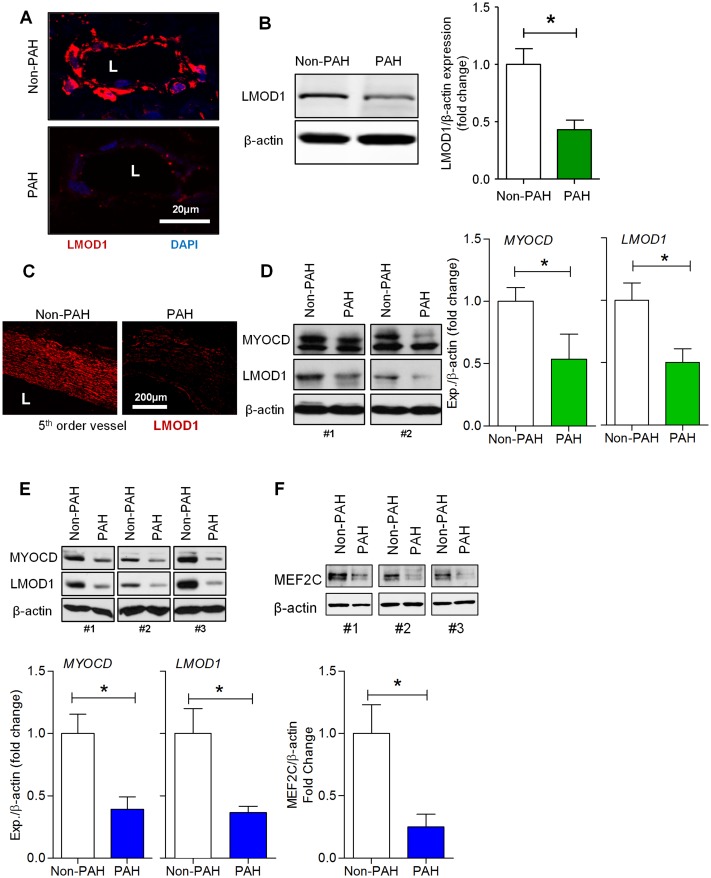
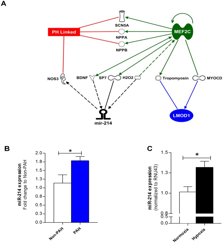
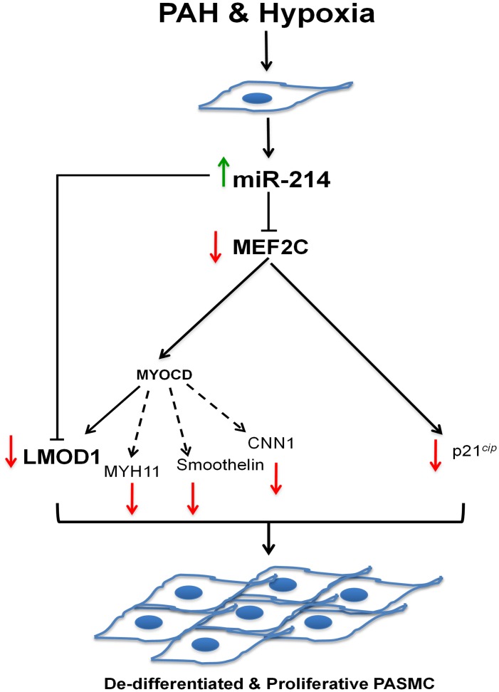
Background: Vascular hyperproliferative disorders are characterized by excessive smooth muscle cell (SMC) proliferation leading to vessel remodeling and occlusion. In pulmonary arterial hypertension (PAH), SMC phenotype switching from a terminally differentiated contractile to synthetic state is gaining traction as our understanding of the disease progression improves. While maintenance of SMC contractile phenotype is reportedly orchestrated by a MEF2C-myocardin (MYOCD) interplay, little is known regarding molecular control at this nexus. Moreover, the burgeoning interest in microRNAs (miRs) provides the basis for exploring their modulation of MEF2C-MYOCD signaling, and in turn, a pro-proliferative, synthetic SMC phenotype. We hypothesized that suppression of SMC contractile phenotype in pulmonary hypertension is mediated by miR-214 via repression of the MEF2C-MYOCD-leiomodin1 (LMOD1) signaling axis.

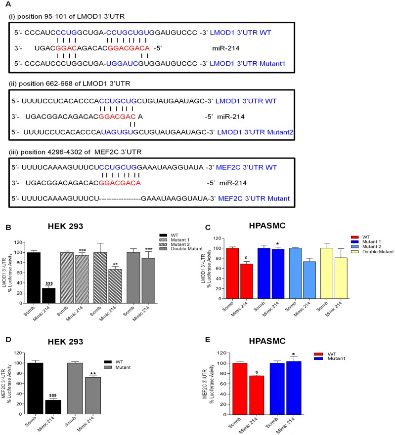
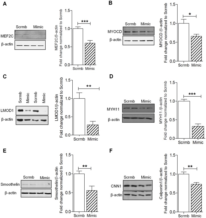
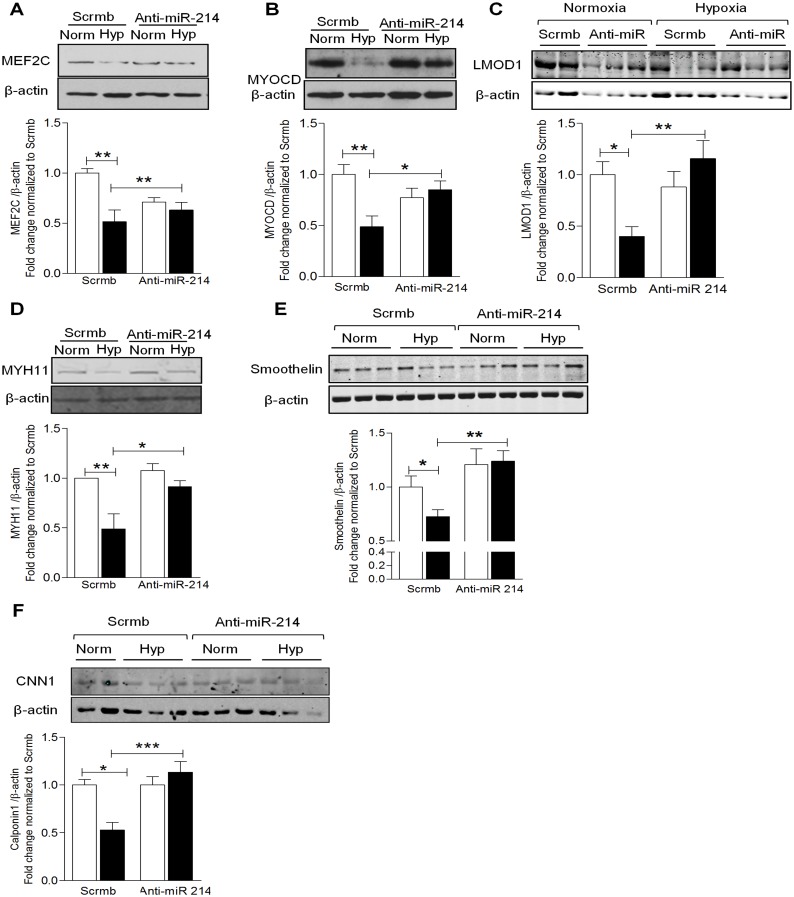
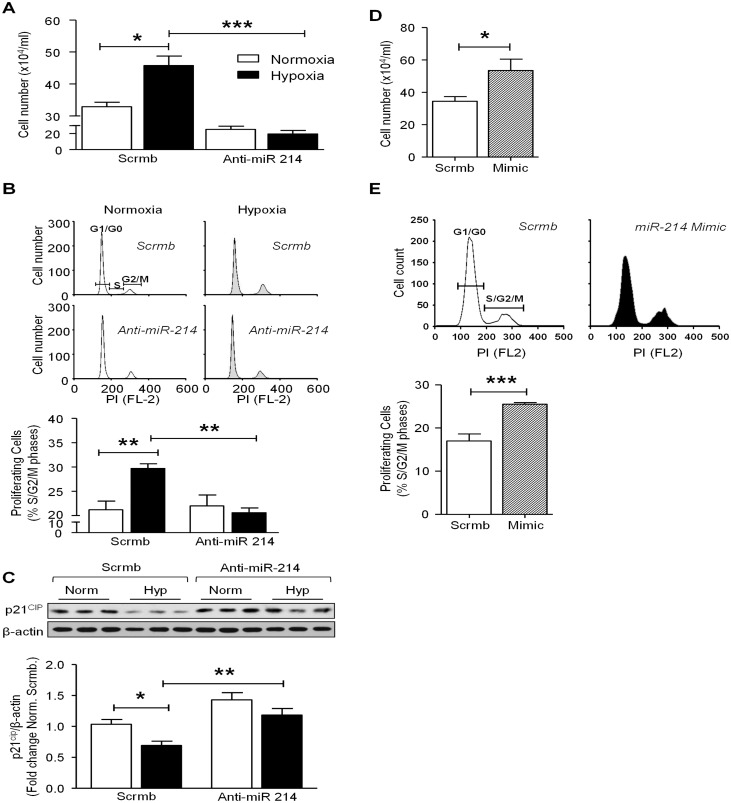
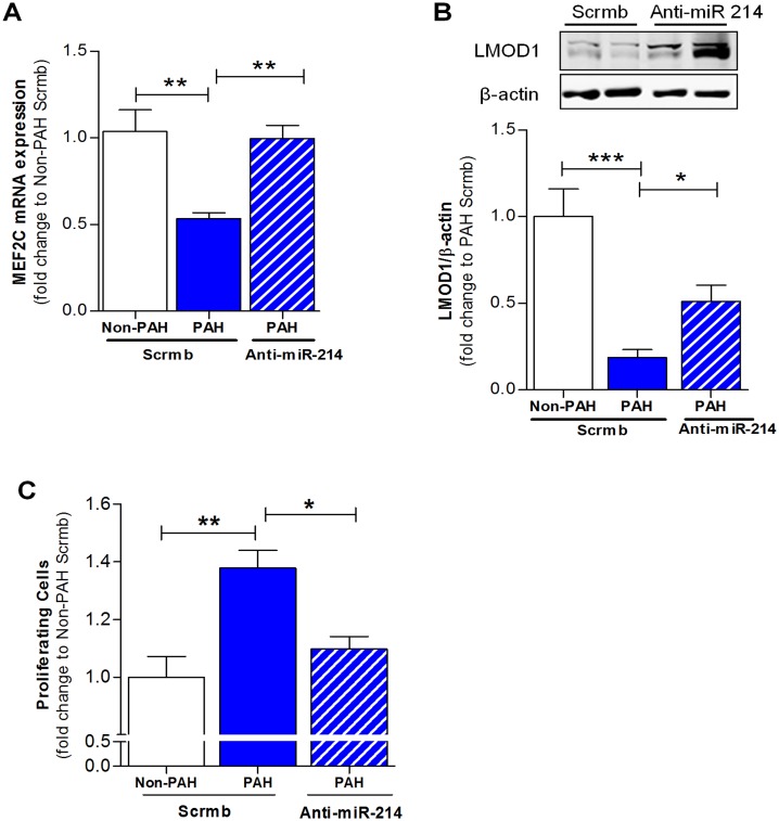
Methods and results: In SMCs isolated from a PAH patient cohort and commercially obtained hPASMCs exposed to hypoxia, miR-214 expression was monitored by qRT-PCR. miR-214 was upregulated in PAH- vs. control subject hPASMCs as well as in commercially obtained hPASMCs exposed to hypoxia. These increases in miR-214 were paralleled by MEF2C, MYOCD and SMC contractile protein downregulation. Of these, LMOD1 and MEF2C were directly targeted by the miR. Mir-214 overexpression mimicked the PAH profile, downregulating MEF2C and LMOD1. AntagomiR-214 abrogated hypoxia-induced suppression of the contractile phenotype and its attendant proliferation. Anti-miR-214 also restored PAH-PASMCs to a contractile phenotype seen during vascular homeostasis.

Conclusions: Our findings illustrate a key role for miR-214 in modulation of MEF2C-MYOCD-LMOD1 signaling and suggest that an antagonist of miR-214 could mitigate SMC phenotype changes and proliferation in vascular hyperproliferative disorders including PAH.


Conflict of interest statement

Competing Interests: The authors have declared that no competing interests exist.

Figures

Fig 1
Fig 1. Contractile proteins, leiomodin1 (LMOD1), myocardin (MYOCD) and MEF2C are downregulated in pulmonary artery smooth muscle cells (hPASMCs) in pulmonary arterial hypertension (PAH).
(A,B) LMOD1 expression in lungs from PAH and non-PAH patients (n = 3) was determined by immunofluorescence and Western blot. Images from lung sections showed a marked decrease in LMOD1 expression (red fluorescence) in the vessel wall, primarily the media, of PAH samples compared to non-PAH group; DAPI nuclear staining (blue), (A). Western blot analysis of total lung homogenates showed a significant decrease in LMOD1 expression in PAH compared to control lung homogenates (B). (C-D) LMOD1 expression in the pulmonary artery (PA) from PAH and non-PAH patients (n = 3) was determined using immunofluorescence and Western blot analysis, respectively. Immunofluorescence images from PA sections of non-PAH samples showed expression of LMOD1 specific to the medial layer (smooth muscle layer, red fluorescence). LMOD1 expression was significantly attenuated in PAH samples compared to non-PAH group (C). Western blot analysis of total PA homogenates showed significant decreases in myocardin (MYOCD) and LMOD1 expression in PAH compared to non-PAH tissue homogenates (D). Western blot analysis of PASMCs-derived from PAH and non-PAH subjects showed decreased levels of MYOCD and LMOD1 protein in PAH group compared to non-PAH group (n = 3–5) (E). Western blot analysis of PASMCs-derived from PAH and non-PAH patients showed decreased levels of MEF2C protein in the PAH group compared to non-PAH subjects (n = 4) (F). Blots are representative; graphs depict mean ± SEM (*, p<0.05).
Fig 2
Fig 2. miR-214 is significantly upregulated in PAH.
(A) Integrated Pathway Analysis (IPA) shows potential association between miR-214-MEF2C-LMOD1 in PAH, and other genes unique to PAH potentially associated with miR-214. Relationships between molecules are represented as follows: bold line, direct interaction; dotted line, indirect interaction; line with arrowhead, represents directionality of interaction; SCN5A, sodium channel, voltage gated, type V alpha subunit; NPPA, natriuretic peptide A; NPPB, natriuretic peptide B; NOS3, nitric oxide synthase, endothelial; BDNF, brain-derived neutrotrophic factor; SP7, Sp7 transcription factor; H2O2, hydrogen peroxide; MEF2C, myocyte enhancer factor 2C; MYOCD, myocardin; LMOD1, leiomodin1. (B) miR-214 expression is increased (~1.6-fold) in SMC-derived from PAH patients compared to controls subjects as measured by q-RT PCR (n = 7–8). (C) Serum-deprived control PASMCs were exposed to normoxia (21% O2) or hypoxia (1% O2) for 24 hrs and, miR-214 expression determined by q-RT PCR. Hypoxia resulted in significant increase in miR-214 expression in PASMCs compared to normoxic controls (n = 8). Graphs represent mean ± SEM. *, p<0.05 versus respective control.
Fig 3
Fig 3. LMOD1 and MEF2C are validated as a direct target of miR-214.
(A) miR-214 binding sites in the 3’-UTR of LMOD1 and MEF2C transcripts. Seed and target sequences are in blue, and base-pairing between miR-214 and target site marked by vertical lines. Mutations in the reporter plasmid construct are indicated in red letters. The double mutant (not shown) carries mutations to both sites in the 3’UTR construct vs. the single site mutants shown in 3A. (B-E) For luciferase assays, HEK293 cells (B, D) or hPASMCs (C, E) were transfected with miR-214 mimic or non-targeting mimic control and wildtype vs. mutant constructs for LMOD1 (B, C) or MEF2C (D, E). Relative luciferase activities were determined 48 hrs post transfection. (n = 3–5), $, p<0.05 WT scrmb vs. mimic; *, p<0.05 WT mimic vs. mutants mimic.
Fig 4
Fig 4. miR-214 regulates expression of SMC contractile genes.
HPASMCs were transfected with miR-214 (Mimic) or scrambled control (Scrmb) under normoxic conditions for 72 hrs and lysed for Western blot analysis. Overexpression of miR-214 using its mimic significantly decreased the expression of MEF2C (A), MYOCD (B), LMOD1 (C), MYH11 (D), smoothelin (E), CNN1 (F), (n = 4–9). Graphs represent mean ± SEM (*, p<0.05, **, p<0.01, ***, p<0.001).
Fig 5
Fig 5. Inhibition of miR-214 restores expression of SMC contractile proteins.
HPASMCs transfected with anti-miR-214 or scrambled control (Scrmb) were exposed to normoxia (open bars) or hypoxia (filled bars) for 24 hrs and lysed for Western blot. Hypoxia significantly decreased the expression of contractile proteins, while transfection with anti-miR-214 significantly reversed the hypoxia-induced suppression of contractile proteins, MEF2C (A), MYOCD (B), LMOD1 (C), MYH11(D), smoothelin (E) and CNN1 (F), (n = 3–6). Graphs represent mean ± SEM (*, p<0.05, **, p<0.01, ***, p<0.001).
Fig 6
Fig 6. miR-214 regulates hypoxia-induced proliferation and cell cycle progression of hPASMCs.
HPASMCs transfected with anti-miR-214 or scrambled control (Scrmb) were exposed to normoxia (open bars) or hypoxia (filled bars) for 24 hrs and harvested for cell counting by trypan blue exclusion (A), trypsinized for flow cytometry analysis using propidium iodide staining (B), or lysed for Western blot analysis (C). In both cell count and cell cycle analysis, hypoxia significantly increased the number of proliferating cells and hPASMC transfected with anti-miR-214 displayed significantly attenuated hypoxia-induced proliferation (n = 5–6). Also, hPASMC transfected with anti-miR-214 attenuated hypoxia-induced increase in S+ G2/M phase cells as depicted by cell cycle analysis. (C) Hypoxia significantly suppressed p21cip levels in hPASMC and anti-miR-214 significantly reversed hypoxia-induced suppression of p21cip. HPASMCs were transfected with miR-214 mimic or scrambled control under normoxic conditions for 72 hrs and harvested for cell counting by trypan blue exclusion (D), trypsinized for flow cytometry analysis using propidium iodide staining (E). miR-214 mimic significantly increased the number of proliferating cells and increased the number of cells in S+ G2/M phase (D, E, respectively; n = 5–6). Graphs represent mean ± SEM (*, p<0.05, **, p<0.01, ***, p<0.001).
Fig 7
Fig 7. miR-214 promotes suppression of MEF2C-LMOD1 signaling axis in PAH-PASMCs.
Analysis of SMC phenotypic markers in PASMCs derived from PAH patients (n = 3–5). PAH-PASMCs were transfected with anti-miR-214 or scrambled control (Scrmb) and lysed for q-RT PCR and Western blot, 72 hrs after transfection. miR-214 antagonism in PAH-PASMCs reversed the expression of MEF2C (A) and LMOD1 (B) comparable to non-PAH controls. (C) Analysis of proliferation in PASMCs derived from PAH patients (n = 3). PAH-PASMCs were transfected with anti-miR-214 or Scrmb and trypsinized for cell cycle analysis 72 hrs after transfection. miR-214 antagonism in PAH-PASMCs significantly inhibited PASMC proliferation. Graphs represent mean ± SEM (*, p<0.05, **, p<0.01, ***, p<0.001).
Fig 8
Fig 8. Schematic of miR-214-mediated effects on PAH-related and hypoxia-induced SMC phenotypic modulation and de-differentiation.
During vascular homeostasis, SMCs maintain a contractile/differentiated phenotype, which changes to a pro-proliferative, synthetic or de-differentiated phenotype upon injury. Hypoxia-induced upregulation of miR-214 exerts direct suppression both at the level of MEF2C and LMOD1 to mediate phenotype switching and pro-proliferative response associated with vascular remodeling.

References

    1. Stenmark KR, Fagan KA, Frid MG (2006) Hypoxia-induced pulmonary vascular remodeling: cellular and molecular mechanisms. Circ Res 99: 675–691. - PubMed
    1. Mandegar M, Fung YC, Huang W, Remillard CV, Rubin LJ, Yuan JX (2004) Cellular and molecular mechanisms of pulmonary vascular remodeling: role in the development of pulmonary hypertension. Microvasc Res 68: 75–103. - PubMed
    1. Rabinovitch M (2007) Pathobiology of pulmonary hypertension. Annu Rev Pathol 2: 369–399. - PubMed
    1. Zhou W, Negash S, Liu J, Raj JU (2009) Modulation of pulmonary vascular smooth muscle cell phenotype in hypoxia: role of cGMP-dependent protein kinase and myocardin. Am J Physiol Lung Cell Mol Physiol 296: L780–789. 10.1152/ajplung.90295.2008 - DOI - PMC - PubMed
    1. Lanner MC, Raper M, Pratt WM, Rhoades RA (2005) Heterotrimeric G proteins and the platelet-derived growth factor receptor-beta contribute to hypoxic proliferation of smooth muscle cells. Am J Respir Cell Mol Biol 33: 412–419. - PubMed

Publication types

MeSH terms