. 2016 Aug;245(8):854-73.
doi: 10.1002/dvdy.24413. Epub 2016 May 31.

CUG-BP, Elav-like family member 1 (CELF1) is required for normal myofibrillogenesis, morphogenesis, and contractile function in the embryonic heart

Affiliations

CUG-BP, Elav-like family member 1 (CELF1) is required for normal myofibrillogenesis, morphogenesis, and contractile function in the embryonic heart

Yotam Blech-Hermoni et al. Dev Dyn. 2016 Aug.

Abstract

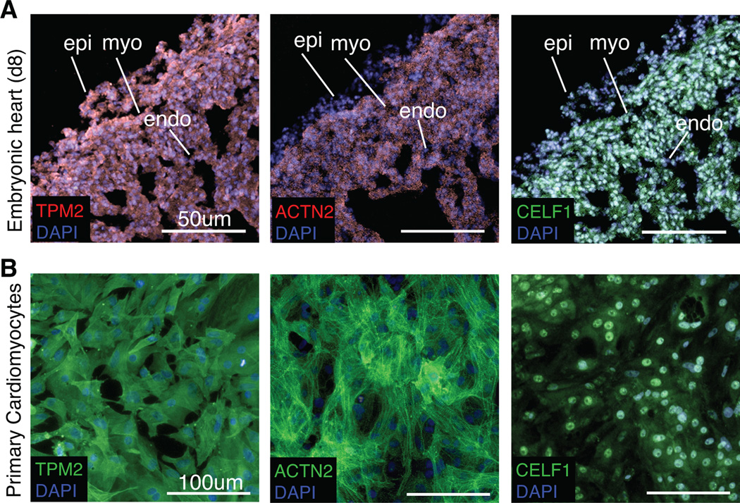
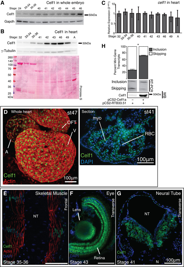
Background: CUG-BP, Elav-like family member 1 (CELF1) is a multifunctional RNA binding protein found in a variety of adult and embryonic tissues. In the heart, CELF1 is found exclusively in the myocardium. However, the roles of CELF1 during cardiac development have not been completely elucidated.

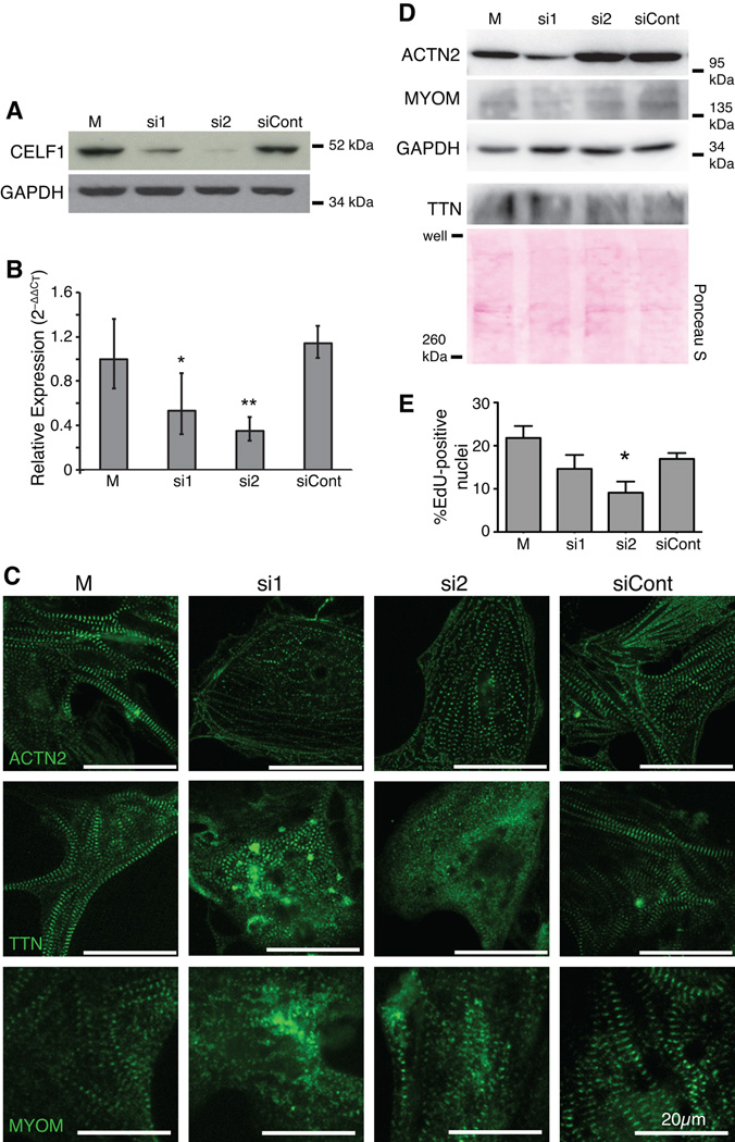
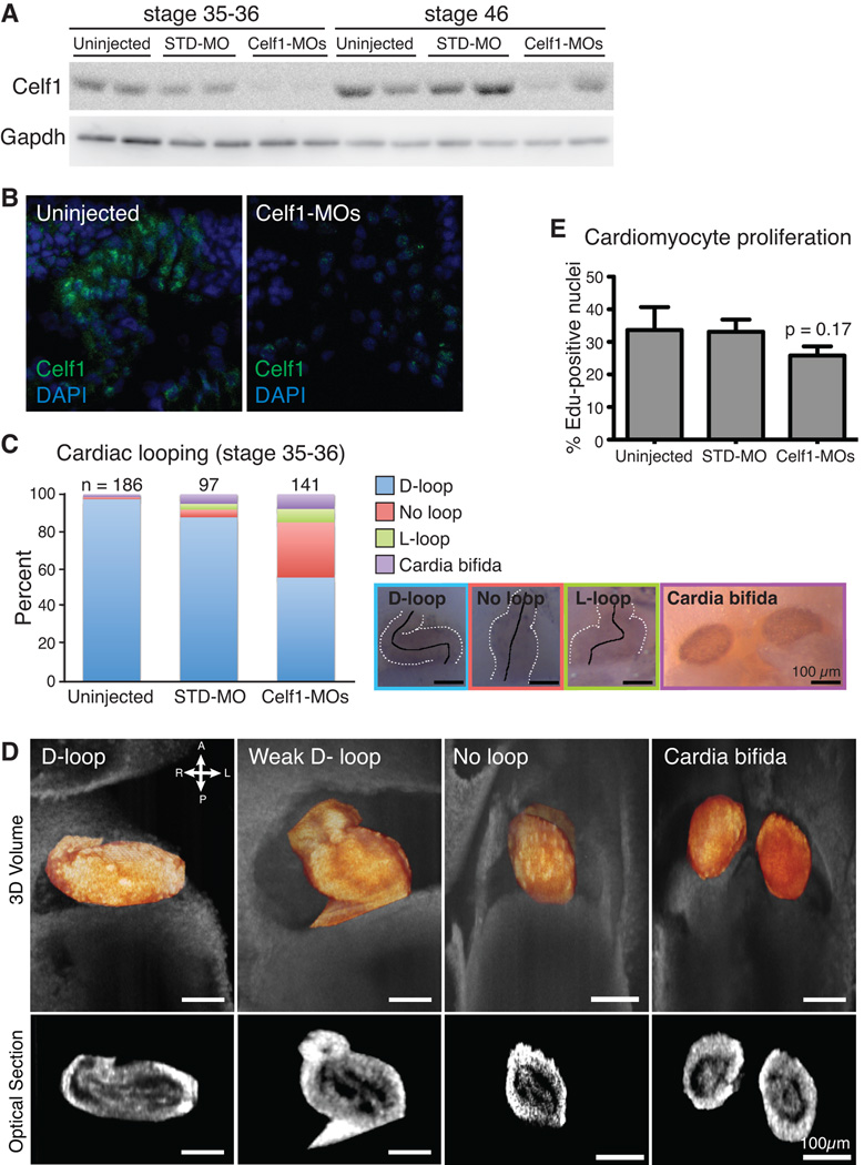
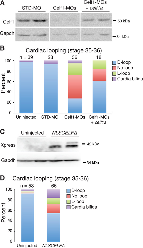
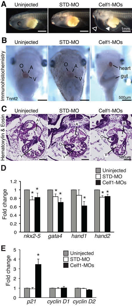
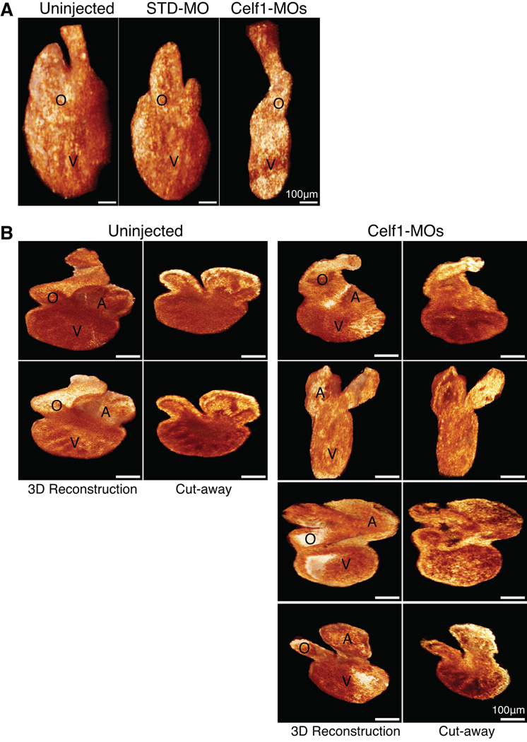
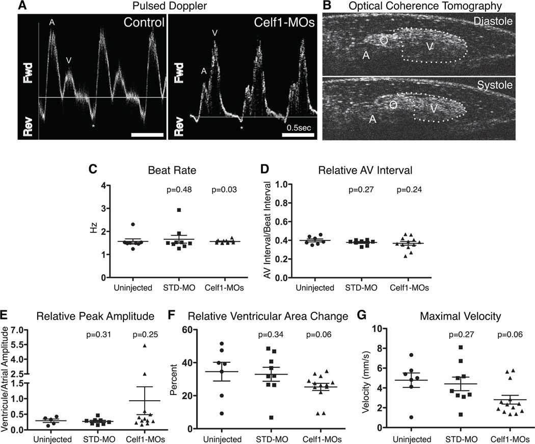
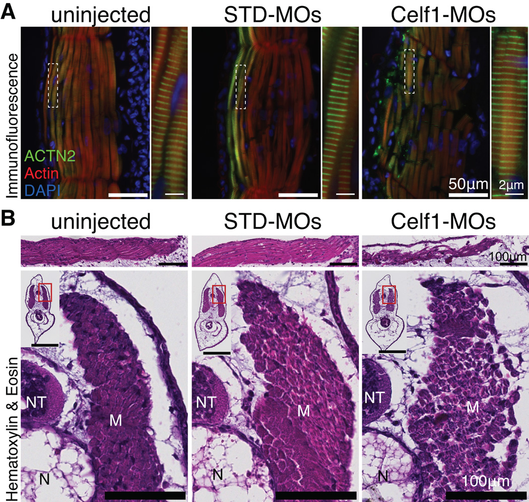
Results: Myofibrillar organization is disrupted and proliferation is reduced following knockdown of CELF1 in cultured chicken primary embryonic cardiomyocytes. In vivo knockdown of Celf1 in developing Xenopus laevis embryos resulted in myofibrillar disorganization and a trend toward reduced proliferation in heart muscle, indicating conserved roles for CELF1 orthologs in embryonic cardiomyocytes. Loss of Celf1 also resulted in morphogenetic abnormalities in the developing heart and gut. Using optical coherence tomography, we showed that cardiac contraction was impaired following depletion of Celf1, while heart rhythm remained unperturbed. In contrast to cardiac muscle, loss of Celf1 did not disrupt myofibril organization in skeletal muscle cells, although it did lead to fragmentation of skeletal muscle bundles.

Conclusions: CELF1 is required for normal myofibril organization, proliferation, morphogenesis, and contractile performance in the developing myocardium. Developmental Dynamics 245:854-873, 2016. © 2016 Wiley Periodicals, Inc.

Keywords: CELF1; Xenopus; chicken; development; heart; myofibrillogenesis; optical coherence tomography.


Figures

Fig. 1
Fig. 1. Cultured chicken primary embryonic cardiomyocytes recapitulate in vivo expression of myofibrillar proteins and CELF1
Cardiomyocytes in the embryonic day 8 chicken heart in vivo (A) and in cultured chicken primary embryonic cardiomyocytes isolated from day 8 hearts (B) express Tropomyosin (TPM2), sarcomeric alpha-Actinin (ACTN2), and CELF1 as determined by immunofluorescence. epi, epicardium; myo, myocardium; endo, endocardium.
Fig. 2
Fig. 2. Knockdown of CELF1 leads to disorganization of myofibrillar structure and reduced proliferation in primary embryonic cardiomyocytes
(A) Depletion of CELF1 with two independent siRNAs (si1 and si2) but not a control siRNA (siCont) is seen by western blot at 72 hours post-transfection; M, mock transfection. Equivalent loading was confirmed by reprobing for GAPDH. (B) Transfection of cells with si1 and si2, but not siCont, resulted in robust knockdown of CELF1 transcripts as measured by real-time RT-PCR. Values shown are means ± 95% confidence intervals. *, p ≤ 0.05; **, p ≤ 0.01 versus mock-transfected cardiomyocytes as evaluated by Student’s t-test. (C) Knockdown of CELF1 at 24 hrs post-plating leads to disruption of myofibril organization as visualized by ACTN2, TTN, and MYOM immunofluorescence at 72 hrs post-transfection. (D) Western blots for ACTN2, TTN, and MYOM protein were performed on mock-, si1-, si2-, and siCont-transfected cardiomyocytes at 72 hrs post-transfection. Protein integrity, equivalent loading, and successful transfer were confirmed by reprobing for GAPDH or Ponceau S staining. (E) Proliferation was measured at 48 hrs post-transfection by EdU incorporation. Values shown are means + standard errors of the mean. *, p ≤ 0.05 versus siCont as evaluated by Student’s t-test.
Fig. 3
Fig. 3. CELF1 is required for myofibril assembly and maintenance
(A) Western blot analysis shows that knockdown of CELF1 protein is detectable by 24 hrs post-transfection (hpt) and persists through 96 hpt. Protein integrity and equivalent loading were confirmed by Ponceau S staining (not shown) and reprobing for GAPDH. (B) Myofibrillar organization was monitored by immunofluorescence using sarcomeric markers (ACTN2, TTN, and MYOM) over 96 hrs following mock (M) or siRNA (si2) transfection. Cells were transfected at 24 hrs post-plating and fixed at 24, 48, 72, and 96 hrs post-transfection; panels match the time points indicated in (A).
Fig. 4
Fig. 4. Developmental expression and alternative splicing activity of Celf1 are conserved in Xenopus laevis
Celf1 protein was evaluated in whole embryo extracts (A) and isolated hearts (B) by western blot analysis. Blots were stripped and reprobed for Gapdh or γ-Tubulin. Protein integrity and equivalent loading were also confirmed by Ponceau S staining. (C) celf1 transcript levels were evaluated in embryonic and adult hearts (n = 3–4) by real-time RT-PCR. Error bars indicate 95% confidence intervals. A one-way analysis of variance found no significant differences in celf1 between the stages. Celf1 was visualized by immunofluorescence in the whole embryonic heart (D, left) and heart sections (D, right), skeletal muscle (E), eye (F), and neural tube (G). A, atrium; V, ventricle; OFT, outflow tract; myo, myocardium; RBC, red blood cell; N, notochord; NT, neural tube. (H) The CELF-responsive RTB33.51 mini-gene was co-expressed in COSM6 cells with celf1-a, RNA was collected 72 hrs later, and alternative splicing was assessed by radiolabeled RT-PCR (top). Celf1 expression was confirmed by western blot analysis (WB). Lanes shown are from the same gels; intervening lanes were removed for clarity. *, p ≤ 0.01; versus mini-gene alone as evaluated by Student’s t-test.
Fig. 5
Fig. 5. Morpholino-mediated Celf1 knockdown in Xenopus laevis causes cardiac looping defects at stage 35–36
(A) Celf1 protein levels were evaluated by western blot analysis (STD-MO, control morpholino; Celf1-MOs, mix of two celf1-targeting morpholinos). (B) Representative images of Celf1 staining in stage 46 heart sections (5–10 sections were stained per embryo for 2 embryos per group from each of 2 injection sets, n = 4 total). Control and Celf1-MOs-injected sections were processed simultaneously and imaged at the same exposure settings and time. STD-MO-injected embryos looked similar to uninjected controls (not shown). (C) Cardiac looping and cardia bifida were evaluated at stage 35–36. Hearts are outlined with broken white lines; a solid black line indicates the middle of the heart tube. D-loop, dextro-loop; L-loop, levo-loop. (D) Tissue overlaying the heart was removed and hearts were imaged in situ by optical coherence tomography (OCT). Three-dimensional reconstructions (top; heart in orange) and optical sections (bottom) are shown. (E) The percentage of cardiomyocytes with EdU-positive nuclei was determined. Data are means + the standard errors of the means of 5–6 embryos from 2–3 injection sets. Pairwise comparisons of the means were performed via Student’s t-test; p value shown for Celf1-MOs-injected group is versus uninjected.
Fig. 6
Fig. 6. Morpholino-mediated knockdown of Celf1 can be ameliorated by restoration of Celf1 expression, and mimicked by repression of nuclear Celf activity
Xenopus laevis embryos were injected either with morpholino oligonucleotides (STD-MO, control morpholino; Celf1-MOs, a mix of two celf1-targeting morpholinos) ± celf1a RNA (A, B), or with RNA encoding a dominant negative CELF protein (NLSCELFΔ; C, D) at the 2-4-cell stage and compared to uninjected controls at stage 35–36. (A) Celf1 protein levels were determined by western blotting. Two embryos per group are shown. Lanes shown are from the same blot. (B) Cardiac looping and the incidence of cardia bifida following knockdown and restoration of Celf1 were evaluated by whole-mount immunohistochemistry using antibodies against meromyosin (MF20) or Tnnt2 (CT3). (C) Dominant negative protein expression was confirmed at stage 26 by western blotting for its Xpress epitope tag. Two embryos per group are shown. (D) Cardiac looping and fusion defects were evaluated in embryos expressing the dominant negative CELF protein by whole mount immunohistochemistry against meromyosin. D-loop, dextro-loop; L-loop, levo-loop.
Fig. 7
Fig. 7. Morpholino-mediated knockdown of Celf1 in Xenopus laevis leads to ventral edema, gut malformation, and cardiac dysmorphia at stage 46
Embryos were injected with morpholino oligonucleotides at the 2-4-cell stage and evaluated at stage 46 (STD-MO, control morpholino; Celf1-MOs, a mix of celf1a- and celf1b-targeting morpholinos). (A) Following depletion of Celf1, most Celf1-MOs-injected embryos exhibited ventral edema (open arrowhead) and many had a malformed gut (filled arrowhead). (B) In embryos stained for Tnnt2, both cardiac and gut dysmorphia can be seen, as well as the aberrant orientation of the heart within the Celf1-MOs-injected embryos. (C) The heart was evaluated by hematoxylin and eosin staining of frontal sections. Serial sections from five embryos per group taken from three separate injection sets were evaluated with similar results. Sections were chosen for similar planes through the heart, since both gross morphology and heart orientation were abnormal in Celf1-MOs-injected embryos. A, atrium; O, outflow tract; V, ventricle. Real-time RT-PCR was performed on RNA from hearts isolated at stage 46 for several cardiac transcription factors (D) and cell cycle genes (E) following Celf1 knockdown (n = 6–7). Error bars indicate standard error of the mean. *, p ≤ 0.05; versus uninjected; Student’s t-test.
Fig. 8
Fig. 8. Cardiac dysmorphia following morpholino-mediated knockdown of Celf1 in Xenopus laevis was imaged in situ and ex vivo at stage 46
The hearts of uninjected (n = 9), STD-MO-injected (n = 7), and Celf1-MOs-injected (n = 8) embryos at stage 46 were imaged in situ by optical coherence tomography (OCT), and then removed and imaged by optical coherence microscopy (OCM). (A) Representative examples of three-dimensional reconstructions of OCT sections through the hearts are shown. General dysmorphia of the heart and outflow tract can be seen following Celf1 depletion; the atria cannot be resolved in these reconstructions. (B) Representative examples of hearts imaged ex vivo using OCM, in intact three-dimensional reconstructions (left) and in cut-away views (right). Multiple hearts from Celf1-MOs-injected embryos are presented to demonstrate the range of dysmorphia observed. Heart reconstructions are shown at equivalent orientations for the purpose of comparison; this orientation does not necessarily match the orientation of the hearts in situ. A, atrium; O, outflow tract; V, ventricle.
Fig. 9
Fig. 9. Morpholino-mediated knockdown of Celf1 in Xenopus laevis leads to myofibril disorganization in the developing heart
Myofibrillar organization was evaluated in heart wall of stage 46 embryos by immunofluorescence. Sections from eight or more embryos per group from six different injection sets were evaluated with similar results; representative images are shown. In uninjected embryos, mature myofibrils are visible as interdigitating Actn2 and F-Actin staining, whereas Actn2 staining is more diffuse in Celf1-MOs-injected embryos, and does not consistently associate with F-Actin striations in the myofibrils. Myofibrils in STD-MO-injected hearts look similar to those in uninjected embryos (not shown). Top panels show the separate channels for the merged images shown below. F-Actin was detected with fluorophore-conjugated phalloidin.
Fig. 10
Fig. 10. Morpholino-mediated knockdown of Celf1 in Xenopus laevis leads to cardiac dysfunction
Uninjected (n = 8), STD-MO-injected (n = 9), and Celf1-MOs-injected (n = 13) embryos were collected at stage 46, immobilized in methylcellulose gel, and imaged by optical coherence tomography (OCT). Doppler signal was measured at the level of the right aortic branch and both Doppler (not shown) and pulsed Doppler traces (A) were generated for live, beating hearts. Fwd, forward flow (positive Doppler shift); Rev, reverse flow (negative Doppler shift); A, atrial peak; V, ventricular peak; *, regurgitation; horizontal line indicates zero-shift. (B) Video-speed OCT movies were recorded in order to evaluate ventricular contraction. A, Atrium; O, OFT; V, ventricle. (C–G) Contractile parameters were measured and compared to uninjected controls using a one-tailed Mann-Whitney U test. Bars indicate mean ± standard error of the mean. Two control embryos that exhibited a single peak on pulsed Doppler were excluded from the analyses in panels (D) and (E).
Fig. 11
Fig. 11. Morpholino-mediated knockdown of Celf1 in Xenopus laevis leads to disorganization of muscle fibers, but not sarcomeric structure, in skeletal muscle
Embryonic skeletal muscle was evaluated in tails of stage 46 embryos (STD-MO, control morpholino, Celf1-MOs, a mix of two celf1-targeting morpholinos). (A) Representative images of frontal sections stained for Actn2, F-Actin, and DAPI by immunofluorescence are shown (n = 4). In uninjected and STD-MO-injected embryos, muscle bundles are tightly organized, while in Celf1-MOs-injected embryos, muscle bundles exhibit fragmentation and loss of cohesion. Regions indicated with dashed boxes are enlarged in adjoining panels. (B) Representative hematoxylin and eosin-stained frontal (top) and transverse (bottom) sections through stage 46 tails are shown. While the appearance of individual muscle fibers was similar in uninjected, STD-MO-injected, and Celf1-MOs-injected embryos, muscle bundles were looser and more fragmented in tails of Celf1-MOs-injected embryos. M, muscle; NT, neural tube; N, notochord. Insets show whole-section views (scale bar = 100 µm); enlarged regions indicated by red boxes.

Similar articles

Cited by

References

    1. Baldwin BR, Timchenko NA, Zahnow CA. Epidermal growth factor receptor stimulation activates the RNA binding protein CUG-BP1 and increases expression of C/EBPbeta-LIP in mammary epithelial cells. Mol Cell Biol. 2004;24:3682–3691. - PMC - PubMed
    1. Barreau C, Paillard L, Mereau A, Osborne HB. Mammalian CELF/Bruno-like RNA-binding proteins: molecular characteristics and biological functions. Biochimie. 2006;88:515–525. - PubMed
    1. Bartlett H, Veenstra GJ, Weeks DL. Examining the cardiac NK-2 genes in early heart development. Pediat Cardiol. 2010;31:335–341. - PMC - PubMed
    1. Berger DS, Ladd AN. Repression of nuclear CELF activity can rescue CELF-regulated alternative splicing defects in skeletal muscle models of myotonic dystrophy. PLoS Curr. 2012;4:RRN1305. - PMC - PubMed
    1. Berger DS, Moyer M, Kliment GM, van Lunteren E, Ladd AN. Expression of a dominant negative CELF protein in vivo leads to altered muscle organization, fiber size, and subtype. PLoS One. 2011;6:e19274. - PMC - PubMed

Publication types

LinkOut - more resources