

Brain GABA levels across psychiatric disorders: A systematic literature review and meta-analysis of (1) H-MRS studies
- PMID: 27145016
- PMCID: PMC6867515
- DOI: 10.1002/hbm.23244
Brain GABA levels across psychiatric disorders: A systematic literature review and meta-analysis of (1) H-MRS studies
Abstract
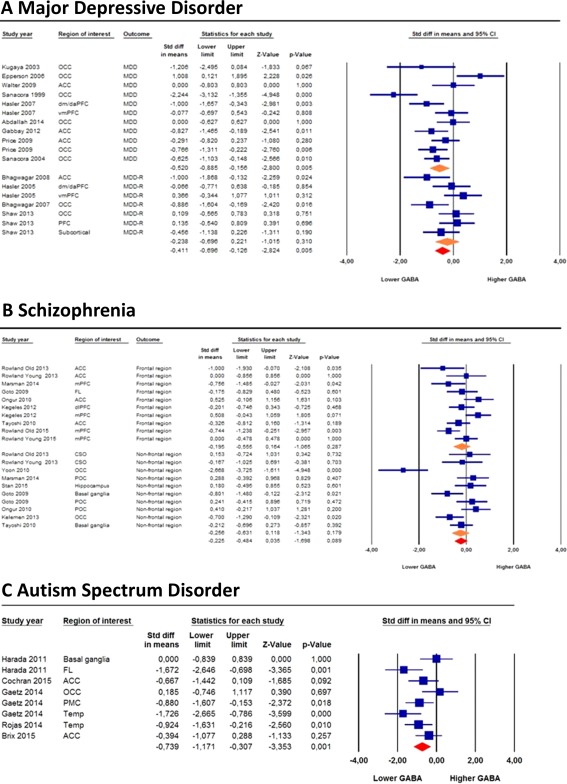
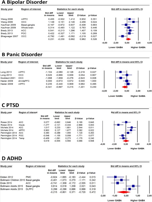
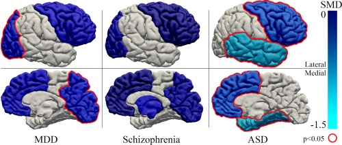
The inhibitory gamma-aminobutyric acid (GABA) system is involved in the etiology of most psychiatric disorders, including schizophrenia, autism spectrum disorder (ASD) and major depressive disorder (MDD). It is therefore not surprising that proton magnetic resonance spectroscopy ((1) H-MRS) is increasingly used to investigate in vivo brain GABA levels. However, integration of the evidence for altered in vivo GABA levels across psychiatric disorders is lacking. We therefore systematically searched the clinical (1) H-MRS literature and performed a meta-analysis. A total of 40 studies (N = 1,591) in seven different psychiatric disorders were included in the meta-analysis: MDD (N = 437), schizophrenia (N = 517), ASD (N = 150), bipolar disorder (N = 129), panic disorder (N = 81), posttraumatic stress disorder (PTSD) (N = 104), and attention deficit/hyperactivity disorder (ADHD) (N = 173). Brain GABA levels were lower in ASD (standardized mean difference [SMD] = -0.74, P = 0.001) and in depressed MDD patients (SMD = -0.52, P = 0.005), but not in remitted MDD patients (SMD = -0.24, P = 0.310) compared with controls. In schizophrenia this finding did not reach statistical significance (SMD = -0.23, P = 0.089). No significant differences in GABA levels were found in bipolar disorder, panic disorder, PTSD, and ADHD compared with controls. In conclusion, this meta-analysis provided evidence for lower brain GABA levels in ASD and in depressed (but not remitted) MDD patients compared with healthy controls. Findings in schizophrenia were more equivocal. Even though future (1) H-MRS studies could greatly benefit from a longitudinal design and consensus on the preferred analytical approach, it is apparent that (1) H-MRS studies have great potential in advancing our understanding of the role of the GABA system in the pathogenesis of psychiatric disorders. Hum Brain Mapp 37:3337-3352, 2016. © 2016 Wiley Periodicals, Inc.
Keywords: 1H-MRS; ASD; GABA; MDD; meta-analysis; psychopathology.
© 2016 Wiley Periodicals, Inc.
Figures
References
-
- Andreychenko A, Boer VO, Arteaga De Castro CS, Luijten PR, Klomp DWJ (2012): Efficient spectral editing at 7 T: GABA detection with MEGA‐sLASER. Magn Reson Med 68:1018–1025. - PubMed
-
- Arteaga De Castro C, Boer V, Andreychenko A, Wijnen J, Van der Heide U, Luijten P, Klomp D (2013): Improved efficiency on editing MRS of lactate and gamma‐aminobutyric acid by inclusion of frequency offset corrected inversion pulses at high fields. NMR Biomed 26:1213–1219. - PubMed
-
- Aufhaus E, Weber‐Fahr W, Sack M, Tunc‐Skarka N, Oberthuer G, Hoerst M, Meyer‐Lindenberg A, Boettcher U, Ende G (2013): Absence of changes in GABA concentrations with age and gender in the human anterior cingulate cortex: A MEGA‐PRESS study with symmetric editing pulse frequencies for macromolecule suppression. Magn Reson Med 69:317–320. - PubMed
Publication types
MeSH terms
Substances
LinkOut - more resources
Full Text Sources
Other Literature Sources
Medical
