. 2016 Aug;170(8):2002-11.
doi: 10.1002/ajmg.a.37721. Epub 2016 May 4.

A novel de novo dominant negative mutation in DNM1L impairs mitochondrial fission and presents as childhood epileptic encephalopathy

Affiliations

A novel de novo dominant negative mutation in DNM1L impairs mitochondrial fission and presents as childhood epileptic encephalopathy

Jill A Fahrner et al. Am J Med Genet A. 2016 Aug.

Abstract

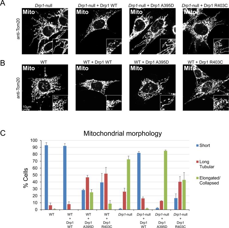
DNM1L encodes dynamin-related protein 1 (DRP1/DLP1), a key component of the mitochondrial fission machinery that is essential for proper functioning of the mammalian brain. Previously reported probands with de novo missense mutations in DNM1L presented in the first year of life with severe encephalopathy and refractory epilepsy, with several dying within the first several weeks after birth. In contrast, we report identical novel missense mutations in DNM1L in two unrelated probands who experienced normal development for several years before presenting with refractory focal status epilepticus and subsequent rapid neurological decline. We expand the phenotype of DNM1L-related mitochondrial fission defects, reveal common unique clinical characteristics and imaging findings, and compare the cellular impact of this novel mutation to the previously reported A395D lethal variant. We demonstrate that our R403C mutation, which resides in the assembly region of DRP1, acts by a dominant-negative mechanism and reduces oligomerization, mitochondrial fission activity, and mitochondrial recruitment of DRP1, but to a lesser extent compared to the A395D mutation. In contrast to the initial report of neonatal lethality resulting from DNM1L mutation and DRP1 dysfunction, our results show that milder DRP1 impairment is compatible with normal early development and subsequently results in a distinct set of neurological findings. In addition, we identify a common pathogenic mechanism whereby DNM1L mutations impair mitochondrial fission. © 2016 Wiley Periodicals, Inc.

Keywords: DLP1; DNM1L; DRP1; developmental regression; epileptic encephalopathy; fission; mitochondria; seizures.


Figures

Figure 1
Figure 1. Magnetic resonance imaging of brains from probands 1 and 2
(A) Axial diffusion-weighted magnetic resonance image with white arrow showing curvilinear intensity in the left thalamus in proband 1. (B,C) Coronal magnetic resonance images obtained from proband 1 at (B) initial presentation and (C) one month following resolution of status epilepticus show progressive atrophy of the brain, most marked in the hippocampi. (D) Axial T2 FLAIR magnetic resonance image with white arrow showing hyperintensity in the right central thalamus in proband 2, which was faintly hyperintense on diffusion-weighted imaging as well. (E,F) Coronal magnetic resonance images obtained from proband 2 at (E) initial presentation and (F) one year following resolution of status epilepticus show progressive atrophy of the brain, particularly involving the right posterior putamen and bilateral hippocampi (left greater than right).
Figure 2
Figure 2. Mutants R403C and A395D have dominant-negative effects on mitochondrial fission
(A) Fission activity upon expression in Drp1-null MEFs. Drp1-null MEFs expressing wild-type Drp1, Drp1A395D, and Drp1R403C were fixed and immunostained against the outer membrane protein TOM20 to visualize mitochondrial morphology. (B) Same as (A) except that wildtype MEFs were used. (E) Quantitation of mitochondrial morphology. Cells were scored into three categories of mitochondrial profiles: short, long tubular, or elongated and/or collapsed mitochondrial tubules. Quantitation was done in triplicate, with 100 cells scored per experiment. Error bars, SEM.
Figure 3
Figure 3. Recruitment to mitochondria is impaired in the A395D and R403C mutants
(A) Analysis of Drp1 alleles in Drp1-null MEFs. Cells expressing the indicated alleles were briefly permeabilized with digitonin to clear cytosolic DRP1 before fixation. This resulted in mitochondrial fragmentation, but DRP1 puncta remained on mitochondria for visualization by immunostaining with an antibody against DRP1 (DRP1, green). Mitochondria were visualized by immunostaining against TOM20 (Mito, red). The DRP1 signal, mitochondrial signal, and merged signals are shown. Inset squares are magnifications of the boxed region in the main image. In the first column, Mff-null MEFs are shown as a reference for defective DRP1 recruitment to mitochondria. (B) Same as (A), except that wildtype MEFs are used.
Figure 4
Figure 4. Assembly defect of DRP1 mutants
(A) DRP1 oligomerization assessed by the yeast two-hybrid assay. DRP1 and DRP1 mutants expressed from the pGAD vector as GAL4 activation domain (AD) fusion proteins were tested against DRP1 expressed from the pGBDU vector as a GAL4 DNA-binding domain (BD) fusion proteins. Growth on adenine-deficient plates indicates an interaction. (B) Ribbon diagram of DRP1 (PDB file: 4BEJ) depicting the location of the R403 and A395 residues. Circled is the region (Interface 3) predicted to mediate dimer-to-dimer interactions during DRP1 oligomerization. (C) Ribbon diagram of the dynamin tetramer (PBD file: 5A3F). The violet and green dynamin monomers comprise the dimer on the left, and the gray and blue monomers comprise the dimer on the right. The inset depicts interface 3 of the dimer:dimer interaction, with the green monomer of the left dimer removed for clarity. The dynamin residues depicted are labeled on the left, next to the corresponding residues on DRP1.

References

    1. Bitoun M, Bevilacqua JA, Prudhon B, Maugenre S, Taratuto AL, Monges S, Lubieniecki F, Cances C, Uro-Coste E, Mayer M, Fardeau M, Romero NB, Guicheney P. Dynamin 2 mutations cause sporadic centronuclear myopathy with neonatal onset. Ann Neurol. 2007;62(6):666–670. - PubMed
    1. Carelli V, Chan DC. Mitochondrial DNA: impacting central and peripheral nervous systems. Neuron. 2014;84(6):1126–1142. - PMC - PubMed
    1. Chan DC. Mitochondrial dynamics in disease. N Engl J Med. 2007;356(17):1707–1709. - PubMed
    1. Chan DC. Fusion and fission: interlinked processes critical for mitochondrial health. Annu Rev Genet. 2012;46:265–287. - PubMed
    1. Euro E-RESC, Epilepsy Phenome/Genome P, Epi KC De novo mutations in synaptic transmission genes including DNM1 cause epileptic encephalopathies. Am J Hum Genet. 2014;95(4):360–370. - PMC - PubMed

MeSH terms