. 2016 May 4;18(1):97.
doi: 10.1186/s13075-016-0996-z.

Selective inhibition of tropomyosin-receptor-kinase A (TrkA) reduces pain and joint damage in two rat models of inflammatory arthritis

Affiliations

Selective inhibition of tropomyosin-receptor-kinase A (TrkA) reduces pain and joint damage in two rat models of inflammatory arthritis

Sadaf Ashraf et al. Arthritis Res Ther. .

Abstract

Background: Inflammation is an essential component of arthritis pain. Nerve growth factor (NGF) plays a key role in acute and chronic pain states especially those associated with inflammation. NGF acts through tropomyosin-receptor-kinase A (TrkA). NGF blockade has reduced arthritis pain in clinical trials. We explored the mechanisms within the joint which may contribute to the analgesic effects of NGF by selectively inhibiting TrkA in carrageenan-induced or collagen-induced joint pain behaviour. The goal of the current study was to elucidate whether inflammation is central to the efficacy for NGF blockade.

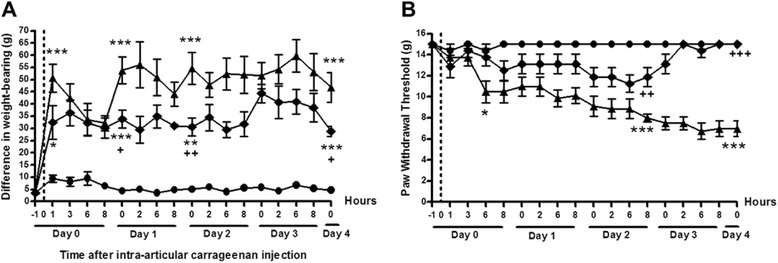
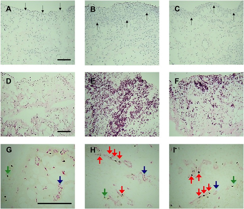
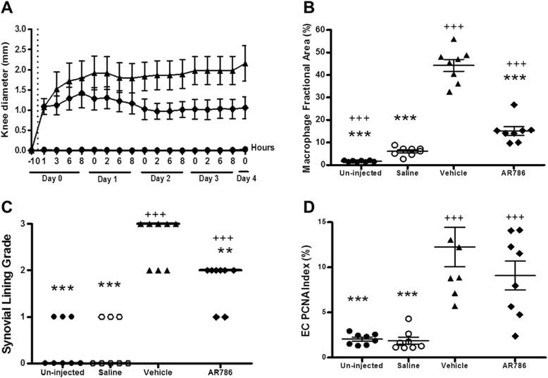
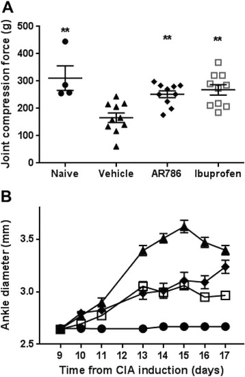
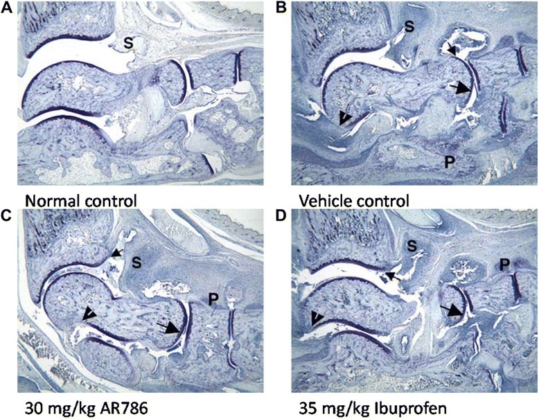
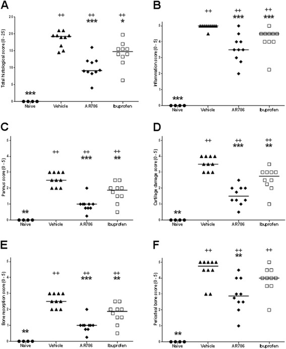
Methods: Rats were injected in their left knees with 2 % carrageenan or saline. Collagen-induced arthritis (CIA) was induced by intradermal injections of a mixture of bovine type II collagen (0.2 mg) and incomplete Freund's adjuvant (0.2 mg). Oral doses (30 mg/kg) of AR786 or vehicle control were given twice daily after arthritis induction. Ibuprofen-treated (35 mg/kg, orally, once daily) rats with CIA were used as positive analgesic controls. Pain behaviour was measured as hind-limb weight-bearing asymmetry and hind-paw withdrawal thresholds to von Frey hair stimulation (carrageenan synovitis), or withdrawal to joint compression using a Randall Selitto device (CIA). Inflammation was measured as increased knee joint diameter and by histopathological analysis.

Results: Intra-articular injections of carrageenan or induction of CIA was each associated with pain behaviour and synovial inflammation. Systemic administration of the TrkA inhibitor AR786 reduced carrageenan-induced or CIA-induced pain behaviour to control values, and inhibited joint swelling and histological evidence of synovial inflammation and joint damage.

Conclusions: By using two models of varying inflammation we demonstrate for the first time that selective inhibition of TrkA may reduce carrageenan-induced or CIA-induced pain behaviour in rats, in part through potentially inhibiting synovial inflammation, although direct effects on sensory nerves are also likely. Our observations suggest that inflammatory arthritis causes pain and the presence of inflammation is fundamental to the beneficial effects (reduction in pain and pathology) of NGF blockade. Further research should determine whether TrkA inhibition may ameliorate human inflammatory arthritis.

Keywords: Carrageenan; Collagen-induced arthritis; Inflammation; Knee; Nerve growth factor; Pain; Tropomyosin-receptor-kinase A.


Figures

Fig. 1
Fig. 1
Effects of the selective tropomyosin-receptor-kinase A (TrkA) inhibitor AR786 on carrageenan-induced pain behaviour. Rat knees were injected with either 2 % carrageenan (triangles and diamonds) or saline (circles) on day 0 (dotted line). Twice-daily oral dose of 30 mg/kg AR786 (diamonds) or 5 % Gelucire vehicle (triangles) control was given 1 h prior to and 8 h after the carrageenan injection and then twice daily (each pair of doses separated by 6 h) through day 4. Vehicle-treated carrageenan-injected animals (triangles) had increased pain behaviour measured as increased difference in weight-bearing (a) and reduced paw withdrawal thresholds (b) through 4 days after carrageenan injection compared with saline-injected controls (circles). Administration AR786 was associated with reduced pain behaviour, with reduced weight-bearing asymmetry from day 1 and increased paw withdrawal thresholds from day 2. Paw withdrawal thresholds were similar in AR786-treated, carrageenan-injected animals to those in saline-injected (non-synovitic) control levels by day 4. Paw withdrawal threshold was not evoked on the contralateral side. *p < 0.05, **p < 0.01 and ***P < 0.001 compared with saline-injected controls; + p < 0.05, ++ p < 0.01, +++ p < 0.001 compared with vehicle-treated, carrageenan-injected animals
Fig. 2
Fig. 2
Effects of selective tropomyosin-receptor-kinase A (TrkA) inhibitor AR786 on carrageenan-induced synovial inflammation and angiogenesis. Saline-treated control animals demonstrate normal synovial lining layer thickness/cellularity (a), macrophage infiltration (d) and endothelial cell (EC) proliferation (g). Four days after intra-articular injection of 2 % carrageenan there was an increase in synovial lining layer thickness/cellularity (b), macrophage infiltration (e) and endothelial cell (EC) proliferation (h). Following treatment with AR786 (c, f, i), synovial lining layer thickness/cellularity (c) and macrophage infiltration (f) were significantly reduced but not to saline control levels (a, d). EC proliferation was not significantly affected following treatment with AR786 (i). Photomicrographs show synovial lining (black arrows) and cellularity as indicated by haematoxylin and eosin staining (ac), macrophages (purple/black) as delineated by immunoreactivity for CD68 (df) and EC (red) as delineated by immunoreactivity for CD31 (blue arrows), proliferating nuclei (black), as delineated by immunoreactivity for proliferating cell nuclear antigen (PCNA) (green arrows), and proliferating ECs (red arrows), which contain PCNA-immunoreactive nuclei (gi). Bars = 100 μm
Fig. 3
Fig. 3
Effects of selective tropomyosin-receptor-kinase A (TrkA) inhibitor AR786 on carrageenan-induced joint inflammation. Rat knees were injected with either 2 % carrageenan (triangles and diamonds) or saline (circles) on day 0 (dotted line). Twice-daily oral doses of 30 mg/kg AR786 (diamonds) or 5 % Gelucire vehicle (triangles) control were given 1 h prior to and 8 h after the carrageenan injection and then twice daily (each pair of doses separated by 6 h) through day 4. Joint swelling (a) in carrageenan-injected knees was partially but significantly reduced following treatment with AR786 (diamonds) when compared to the vehicle-treated carrageenan-injected animals (triangles) (increased AUC over saline-injected, non-inflamed control knees 9.3 (95 % CI 8.4 to 10.1) mm/day versus 15.0 (95 % CI 13.4 to 16.6) mm/day, p < 0.001). Four days after carrageenan injection, macrophage infiltration (b) and synovial lining layer thickness/cellularity (c) were partially reduced, although synovial angiogenesis (endothelial cell (EC) proliferation index) (d) was not significantly affected in rats that were treated with AR786 compared with vehicle-treated, carrageenan-injected controls. **p < 0.01, ***p < 0.001 versus vehicle-treated carrageenan-injected animals; ++ p < 0.01, +++ p < 0.001 versus saline-injected (non-synovitic) controls. Horizontal bars (c) represent median values
Fig. 4
Fig. 4
Effects of selective tropomyosin-receptor-kinase A (TrkA) inhibitor AR786 on pain behaviour and ankle swelling following collagen-induced arthritis (CIA). Rats were sensitised and challenged to type II bovine collagen in incomplete Freund’s adjuvant. Oral doses of 30 mg/kg AR786, twice daily or 35 mg/kg ibuprofen once daily were administered from the day of collagen challenge. a On day 17 pain was measured in the rats using a Randall Selito device. CIA was associated with decreased force required to elicit a withdrawal response. AR786 or ibuprofen each increased the force required to elicit a response to values that did not differ significantly from non-arthritic controls. b AR786 (diamonds) or ibuprofen (open squares) each resulted in significant inhibition of ankle swelling compared to vehicle-treated rats with CIA (triangles) (each p < 0.005), but the effect did not reach control levels (circles) (each p < 0.007) (AUC (mm/day) vehicle-treated non-arthritic control 22.6 (95 % CI 21.5 to 23.6), vehicle-treated CIA 33.4 (95 % CI, 31.9 to 34.8), AR786-treated CIA 28.0 (95 % CI 26.1 to 30.0), ibuprofen-treated CIA 27.0 (95 % CI, 26.0 to 28.1); analysis of variance F = 32.3, p < 0.001. *p < 0.05, **p < 0.01, ***p < 0.001 versus vehicle (5 % Gelucire) controls
Fig. 5
Fig. 5
Histological appearances of ankles from rats with collagen-induced arthritis treated with AR786 or ibuprofen. Untreated control animal (a) displays normal synovium (S), whereas an ankle from a vehicle-treated arthritic animal (b) displays severe synovitis (S) and moderate cartilage damage (large arrow) with mild pannus (small arrow) and bone resorption (arrowhead). P identifies very severe periosteal bone formation. c Ankle from an arthritic animal treated with AR786 has marked synovitis (S) and mild cartilage damage (large arrow) with minimal pannus (small arrow) and bone resorption. P identifies mild periosteal bone formation. d Ankle from an animal treated with 35 mg/kg ibuprofen has severe inflammation (S) and moderate cartilage damage (large arrow) with mild pannus (small arrow) and bone resorption (arrowhead). P identifies moderate periosteal bone formation. Toluidine blue stain
Fig. 6
Fig. 6
Effects of selective tropomyosin-receptor-kinase A (TrkA) inhibitor AR786 on ankle pathology following collagen-induced arthritis. Collagen-induced arthritis was associated with increased total histological score (a), synovial inflammation (b), pannus formation (c), cartilage damage (d), bone resorption (e) and periosteal bone formation (f). AR786 30 mg/kg orally twice daily from the time of collagen challenge was associated with reduced total histological score and each of its components after 17 days. Ibuprofen 35 mg/kg orally once daily also significantly reduced total histological scores, inflammation, pannus, cartilage damage and bone resorption sub-scores. Kruskal-Wallis statistics all >19, p ≤ 0.0002. Comparisons versus vehicle-treated arthritic animals, *p < 0.05, **p < 0.01, ***p < 0.001, and versus naïve controls + p < 0.05, ++ p < 0.01, +++ p < 0.001. Horizontal bars represent median values

Similar articles

Cited by

References

    1. Aloe L, Tuveri MA, Carcassi U, Levi-Montalcini R. Nerve growth factor in the synovial fluid of patients with chronic arthritis. Arthritis Rheum. 1992;35(3):351–5. doi: 10.1002/art.1780350315. - DOI - PubMed
    1. Manni L, Lundeberg T, Fiorito S, Bonini S, Vigneti E, Aloe L. Nerve growth factor release by human synovial fibroblasts prior to and following exposure to tumor necrosis factor-alpha, interleukin-1 beta and cholecystokinin-8: the possible role of NGF in the inflammatory response. Clin Exp Rheumatol. 2003;21(5):617–24. - PubMed
    1. Walsh DA, Bonnet CS, Turner EL, Wilson D, Situ M, McWilliams DF. Angiogenesis in the synovium and at the osteochondral junction in osteoarthritis. Osteoarthritis Cartilage. 2007;15(7):743–51. doi: 10.1016/j.joca.2007.01.020. - DOI - PubMed
    1. Walsh DA, McWilliams DF, Turley MJ, Dixon MR, Franses RE, Mapp PI, et al. Angiogenesis and nerve growth factor at the osteochondral junction in rheumatoid arthritis and osteoarthritis. Rheumatology (Oxford) 2010;49(10):1852–61. doi: 10.1093/rheumatology/keq188. - DOI - PMC - PubMed
    1. Ashraf S, Mapp PI, Walsh DA. Angiogenesis and the persistence of inflammation in a rat model of proliferative synovitis. Arthritis Rheum. 2010;62(7):1890–8. - PubMed