

Mechanisms of Mitotic Spindle Assembly
- PMID: 27145846
- PMCID: PMC5016079
- DOI: 10.1146/annurev-biochem-060815-014528
Mechanisms of Mitotic Spindle Assembly
Abstract
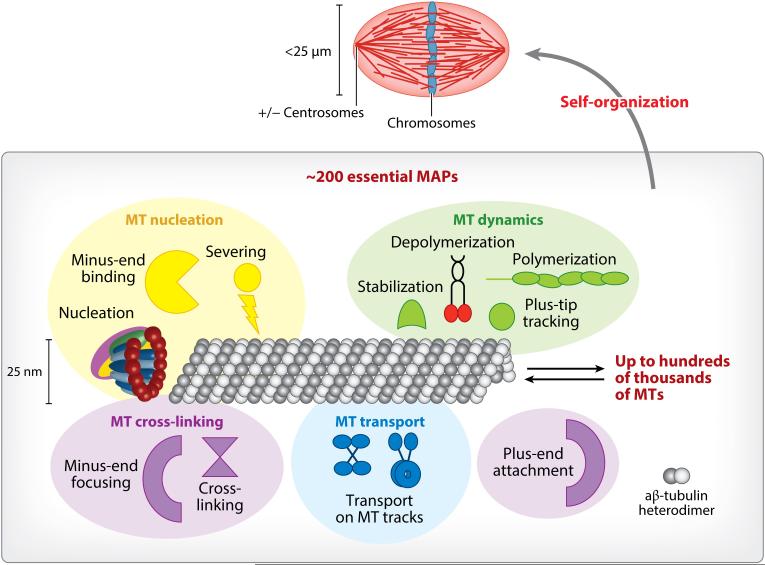
Life depends on cell proliferation and the accurate segregation of chromosomes, which are mediated by the microtubule (MT)-based mitotic spindle and ∼200 essential MT-associated proteins. Yet, a mechanistic understanding of how the mitotic spindle is assembled and achieves chromosome segregation is still missing. This is mostly due to the density of MTs in the spindle, which presumably precludes their direct observation. Recent insight has been gained into the molecular building plan of the metaphase spindle using bulk and single-molecule measurements combined with computational modeling. MT nucleation was uncovered as a key principle of spindle assembly, and mechanistic details about MT nucleation pathways and their coordination are starting to be revealed. Lastly, advances in studying spindle assembly can be applied to address the molecular mechanisms of how the spindle segregates chromosomes.
Keywords: cell division; cytoskeleton; microtubule; microtubule nucleation; microtubule-associated protein; γ-tubulin ring complex.
Figures
Similar articles
-
The microtubule-associated protein EML3 regulates mitotic spindle assembly by recruiting the Augmin complex to spindle microtubules.J Biol Chem. 2019 Apr 5;294(14):5643-5656. doi: 10.1074/jbc.RA118.007164. Epub 2019 Feb 5. J Biol Chem. 2019. PMID: 30723163 Free PMC article.
-
Nudel/NudE and Lis1 promote dynein and dynactin interaction in the context of spindle morphogenesis.Mol Biol Cell. 2013 Nov;24(22):3522-33. doi: 10.1091/mbc.E13-05-0283. Epub 2013 Sep 11. Mol Biol Cell. 2013. PMID: 24025714 Free PMC article.
-
Factors that Control Mitotic Spindle Dynamics.Adv Exp Med Biol. 2017;925:89-101. doi: 10.1007/5584_2016_74. Adv Exp Med Biol. 2017. PMID: 27722958 Review.
-
FAM29A promotes microtubule amplification via recruitment of the NEDD1-gamma-tubulin complex to the mitotic spindle.J Cell Biol. 2008 Dec 1;183(5):835-48. doi: 10.1083/jcb.200807046. Epub 2008 Nov 24. J Cell Biol. 2008. PMID: 19029337 Free PMC article.
-
Cooperative mechanisms of mitotic spindle formation.J Cell Sci. 2007 May 15;120(Pt 10):1717-22. doi: 10.1242/jcs.03442. J Cell Sci. 2007. PMID: 17502482 Review.
Cited by
-
Augmin prevents merotelic attachments by promoting proper arrangement of bridging and kinetochore fibers.Elife. 2022 Oct 21;11:e83287. doi: 10.7554/eLife.83287. Elife. 2022. PMID: 36269126 Free PMC article.
-
SET1/MLL family of proteins: functions beyond histone methylation.Epigenetics. 2021 May;16(5):469-487. doi: 10.1080/15592294.2020.1809873. Epub 2020 Aug 31. Epigenetics. 2021. PMID: 32795105 Free PMC article. Review.
-
Identifying Mitotic Kinesins as Potential Prognostic Biomarkers in Ovarian Cancer Using Bioinformatic Analyses.Diagnostics (Basel). 2022 Feb 12;12(2):470. doi: 10.3390/diagnostics12020470. Diagnostics (Basel). 2022. PMID: 35204562 Free PMC article.
-
Genetic Control of Kinetochore-Driven Microtubule Growth in Drosophila Mitosis.Cells. 2022 Jul 6;11(14):2127. doi: 10.3390/cells11142127. Cells. 2022. PMID: 35883570 Free PMC article.
-
Reconstitution and mechanistic dissection of the human microtubule branching machinery.J Cell Biol. 2022 Jul 4;221(7):e202109053. doi: 10.1083/jcb.202109053. Epub 2022 May 23. J Cell Biol. 2022. PMID: 35604367 Free PMC article.
References
-
- Bianconi E, Piovesan A, Facchin F, Beraudi A, Casadei R, et al. An estimation of the number of cells in the human body. Ann. Hum. Biol. 2013;40(6):463–71. - PubMed
-
- Flemming W. Zellsubstanz, Kern und Zelltheilung. F.C.W. Vogel; Leipzig, Ger.: 1882.
-
- Sauer G, Korner R, Hanisch A, Ries A, Nigg EA, Sillje HH. Proteome analysis of the human mitotic spindle. Mol. Cell. Proteomics. 2005;4:35–43. - PubMed
-
- Brugues J, Nuzzo V, Mazur E, Needleman DJ. Nucleation and transport organize microtubules in metaphase spindles. Cell. 2012;149:554–64. - PubMed
Publication types
MeSH terms
Substances
Grants and funding
LinkOut - more resources
Full Text Sources
Other Literature Sources
