HIV-1 Infection-Induced Suppression of the Let-7i/IL-2 Axis Contributes to CD4(+) T Cell Death
- PMID: 27145859
- PMCID: PMC4857132
- DOI: 10.1038/srep25341
HIV-1 Infection-Induced Suppression of the Let-7i/IL-2 Axis Contributes to CD4(+) T Cell Death
Abstract
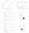
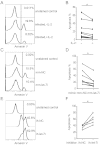
The mechanisms underlying HIV-1-mediated CD4(+) T cell depletion are highly complicated. Interleukin-2 (IL-2) is a key cytokine that maintains the survival and proliferation of activated CD4(+) T cells. IL-2 levels are disturbed during HIV-1 infection, but the underlying mechanism(s) requires further investigation. We have reported that cellular microRNA (miRNA) let-7i upregulates IL-2 expression by targeting the promoter TATA-box region, which functions as a positive regulator. In this study, we found that HIV-1 infection decreases the expression of let-7i in CD4(+) T cells by attenuating its promoter activity. The reduced let-7i miRNA expression led to a decline in IL-2 levels. A let-7i mimic increased IL-2 expression and subsequently enhanced the resistance of CD4(+) T cells to HIV-1-induced apoptosis. By contrast, the blockage of let-7i with a specific inhibitor resulted in elevated CD4(+) T cell apoptosis during HIV-1 infection. Furthermore, by knocking down the expression of IL-2, we found that the let-7i-mediated CD4(+) T cell resistance to apoptosis during HIV-1 infection was dependent on IL-2 signaling rather than an alternative CD95-mediated cell-death pathway. Taken together, our findings reveal a novel pathway for HIV-1-induced dysregulation of IL-2 cytokines and depletion of CD4(+) T-lymphocytes.
Figures


Similar articles
-
RNAa Induced by TATA Box-Targeting MicroRNAs.Adv Exp Med Biol. 2017;983:91-111. doi: 10.1007/978-981-10-4310-9_7. Adv Exp Med Biol. 2017. PMID: 28639194
-
MicroRNA let-7i regulates dendritic cells maturation targeting interleukin-10 via the Janus kinase 1-signal transducer and activator of transcription 3 signal pathway subsequently induces prolonged cardiac allograft survival in rats.J Heart Lung Transplant. 2016 Mar;35(3):378-388. doi: 10.1016/j.healun.2015.10.041. Epub 2015 Nov 22. J Heart Lung Transplant. 2016. PMID: 26755202
-
Differential regulation of the Let-7 family of microRNAs in CD4+ T cells alters IL-10 expression.J Immunol. 2012 Jun 15;188(12):6238-46. doi: 10.4049/jimmunol.1101196. Epub 2012 May 14. J Immunol. 2012. PMID: 22586040
-
Dual mechanism of Let-7i in tumor progression.Front Oncol. 2023 Sep 27;13:1253191. doi: 10.3389/fonc.2023.1253191. eCollection 2023. Front Oncol. 2023. PMID: 37829341 Free PMC article. Review.
-
The role of surface CD4 in HIV-induced apoptosis.Adv Exp Med Biol. 1995;374:91-9. doi: 10.1007/978-1-4615-1995-9_8. Adv Exp Med Biol. 1995. PMID: 7572403 Review. No abstract available.
Cited by
-
Analysis of the Contribution of 6-mer Seed Toxicity to HIV-1-Induced Cytopathicity.J Virol. 2023 Jul 27;97(7):e0065223. doi: 10.1128/jvi.00652-23. Epub 2023 Jun 13. J Virol. 2023. PMID: 37310263 Free PMC article.
-
The Role of MicroRNAs in HIV Infection.Genes (Basel). 2024 Apr 29;15(5):574. doi: 10.3390/genes15050574. Genes (Basel). 2024. PMID: 38790203 Free PMC article. Review.
-
Chronic delta-9-tetrahydrocannabinol (THC) treatment counteracts SIV-induced modulation of proinflammatory microRNA cargo in basal ganglia-derived extracellular vesicles.J Neuroinflammation. 2022 Sep 12;19(1):225. doi: 10.1186/s12974-022-02586-9. J Neuroinflammation. 2022. PMID: 36096938 Free PMC article.
-
Let-7i: A key player and a promising biomarker in diseases.Zhong Nan Da Xue Xue Bao Yi Xue Ban. 2023 Jun 28;48(6):909-919. doi: 10.11817/j.issn.1672-7347.2023.220146. Zhong Nan Da Xue Xue Bao Yi Xue Ban. 2023. PMID: 37587077 Free PMC article. Chinese, English.
-
Expression of microRNAs in the detection and therapeutic roles of viral infections: Mechanisms and applications.J Virus Erad. 2025 Mar 12;11(1):100586. doi: 10.1016/j.jve.2025.100586. eCollection 2025 Mar. J Virus Erad. 2025. PMID: 40296890 Free PMC article. Review.
References
-
- Li C. J., Friedman D. J., Wang C., Metelev V. & Pardee A. B. Induction of apoptosis in uninfected lymphocytes by HIV-1 Tat protein. Science 268, 429–431 (1995). - PubMed
Publication types
MeSH terms
Substances
Grants and funding
LinkOut - more resources
Full Text Sources
Other Literature Sources
Medical
Research Materials

