Review
. 2016 Oct 6:32:373-397.
doi: 10.1146/annurev-cellbio-100814-125227. Epub 2016 May 4.

How Bacteria Subvert Animal Cell Structure and Function

Affiliations
Review

How Bacteria Subvert Animal Cell Structure and Function

Alyssa Jimenez et al. Annu Rev Cell Dev Biol. .

Abstract

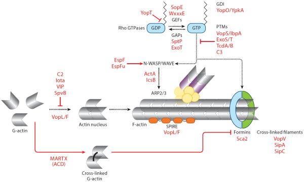
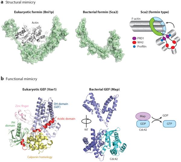
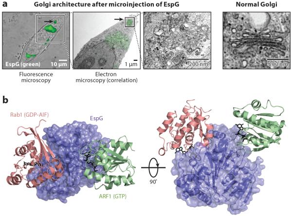
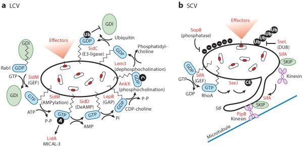
Bacterial pathogens encode a wide variety of effectors and toxins that hijack host cell structure and function. Of particular importance are virulence factors that target actin cytoskeleton dynamics critical for cell shape, stability, motility, phagocytosis, and division. In addition, many bacteria target organelles of the general secretory pathway (e.g., the endoplasmic reticulum and the Golgi complex) and recycling pathways (e.g., the endolysosomal system) to establish and maintain an intracellular replicative niche. Recent research on the biochemistry and structural biology of bacterial effector proteins and toxins has begun to shed light on the molecular underpinnings of these host-pathogen interactions. This exciting work is revealing how pathogens gain control of the complex and dynamic host cellular environments, which impacts our understanding of microbial infectious disease, immunology, and human cell biology.

Keywords: Golgi complex; actin cytoskeleton; bacterial effectors; bacterial toxins; endosomal/lysosomal trafficking.


Figures

Figure 1
Figure 1
Regulation of actin dynamics by bacterial toxins and effector proteins. Bacterial pathogens have evolved multiple strategies to both positively and negatively regulate actin dynamics. Actin polymerization is inhibited through ADP-ribosylation of G-actin into nonpolymerizable monomers by the C2 toxin, the iota toxin, the VIP (vegetative insecticidal protein) toxin, and the Salmonella effector SpvB. ACD (actin cross-linking domain) toxin–mediated actin cross-linking poisons the formin family of actin nucleators. Actin polymerization is inhibited by bacterial effectors through the removal of membrane-bound Rho GTPases via either sequestration of the lipid group by YopO/YpkA or proteolytic cleavage of the lipid group from Rho GTPases by YopT. Additionally, inactivation of Rho GTPases via effector GAP (GTPase-activating protein) activity of SptP and ExoT shuts off actin polymerization. Rho GTPases undergo posttranslational modification (PTM) by ADP-ribosylation via C3 toxins and effectors ExoS and ExoT and by AMPylation by VopS and IbpA, preventing Rho GTPase activation of actin polymerization. Bacterial pathogens have also evolved mechanisms to induce actin dynamics through various mechanisms. VopL/F, Sca2, and SipC directly nucleate actin. Additionally, VopV, SipC, and SipA stabilize actin filaments by cross-linking. Bacterial effectors directly activate or function as nucleation-promoting factors to promote Arp2/3-mediated actin nucleation. Activation of Rho GTPase signaling by SopE- and WxxxE-family members of bacterial GEFs (guanine nucleotide exchange factors) also induces actin polymerization.
Figure 2
Figure 2
Bacterial effectors mimic eukaryotic actin-binding proteins and regulators. Bacterial effector proteins can structurally or functionally mimic host proteins. (a) The Rickettsia actin nucleator Sca2 (PDB: 4J7O) is a structural mimic of eukaryotic formin Bni1p (PDB: 1Y64) (Otomo et al. 2005). Formins adopt a ringlike structure that aids in barbed-end polymerization. (b) Several classes of bacterial guanine nucleotide exchange factors (GEFs), including SopE- and WxxxE-family members (Escherichia coli Map shown here; PDB: 3GCG), functionally mimic eukaryotic GEFs (Vav1 shown here; PDB: 3KY9). Both bacterial and eukaryotic GEFs of the Dbl family induce similar structural rearrangements in Rho GTPase proteins, but they do so using completely distinct structural architectures and catalytic residues. In addition, unlike eukaryotic GEFs, which are highly decorated with various accessory domains, bacterial GEFs are compact and encode minimal accessory domains. Abbreviations: DH, Dbl homology domain; PH, pleckstrin homology domain.
Figure 3
Figure 3
Bacterial effector regulation of organelle structure and function. (a) As shown by correlative light electron microscopy, the Golgi apparatus vesiculates shortly after microinjection of the Escherichia coli effector EspG. A normal Golgi apparatus is shown at the right. (b) The structure of EspG in complex with Rab1 and ARF1 has provided important insights into the mechanism of EspG-mediated Golgi vesiculation (PDB: 4FME).
Figure 4
Figure 4
Effector-mediated remodeling of pathogen-containing vacuoles: Legionella-containing vacuoles (LCV) and Salmonella-containing vacuoles (SCV). (a) Legionella encodes multiple effectors that regulate Rab1 activity. SidM is a multidomain effector with both Rab1 GEF (guanine nucleotide exchange factor) activity and AMPylation activity (black circle labeled A). SidD can deAMPylate Rab1. Rab1 can be inactivated by the GAP (GTPase-activating protein) LepB. Furthermore, Rab1 can be modified by the addition of phosphocholine by AnkX through its FIC (filamentation induced by cAMP) domain (black circle labeled C) or by the removal of the phosphocholine by Lem3. Lastly, the ubiquitin ligase SidC is thought to ubiquitinate (black circle labeled Ub) Rab1, preventing its interaction with its GDI (guanine nucleotide dissociation inhibitor). (b) Salmonella modifies its SCV through the action of multiple effectors. The SPI-1-secreted effector SopB, a phosphoinositide phosphatase, is involved in invasion and alters the surface charge (black circles labeled with minus signs) of the SCV to prevent lysosomal fusion. SifA is a key effector regulating SCV maintenance and Salmonella-induced filament (SIF) formation. SifA, a SPI-2 effector, interacts with the host protein SKIP (SifA kinesin-interacting protein) to retain SCV perinuclear positioning through kinesin and PipB2 interactions. In addition, the GEF domain of SifA is thought to activate RhoA GTPase at the SCV, which subsequently induces the acyltransferase activity of the SPI-2 effector SseJ, generating cholesterol esters on the SCV (black circle labeled CE). These events are thought to promote SIF formation. SseL, through its deubiquitinase (DUB) activity, reverses SCV ubiquitination and host cell recognition by autophagosomes.

References

    1. Aili M, Hallberg B, Wolf-Watz H, Rosqvist R. GAP activity of Yersinia YopE. Methods Enzymol. 2002;358:359–70. - PubMed
    1. Aktories K. Bacterial protein toxins that modify host regulatory GTPases. Nat. Rev. Microbiol. 2011;9:487–98. - PubMed
    1. Aktories K, Barmann M, Ohishi I, Tsuyama S, Jakobs KH, Habermann E. Botulinum C2 toxin ADP-ribosylates actin. Nature. 1986;322:390–92. - PubMed
    1. Aktories K, Wegner A. ADP-ribosylation of actin by clostridial toxins. J. Cell Biol. 1989;109:1385–87. - PMC - PubMed
    1. Al-Younes HM, Brinkmann V, Meyer TF. Interaction of Chlamydia trachomatis serovar L2 with the host autophagic pathway. Infect. Immun. 2004;72:4751–62. - PMC - PubMed

Publication types

LinkOut - more resources