. 2016 May 5:6:25374.
doi: 10.1038/srep25374.

A Novel Protective Function of 5-Methoxytryptophan in Vascular Injury

Affiliations

A Novel Protective Function of 5-Methoxytryptophan in Vascular Injury

Yen-Chun Ho et al. Sci Rep. .

Abstract

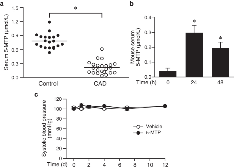
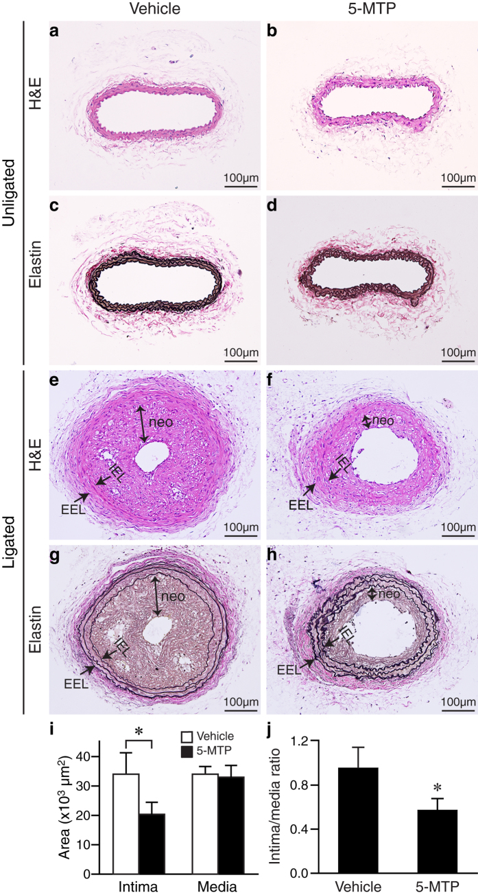
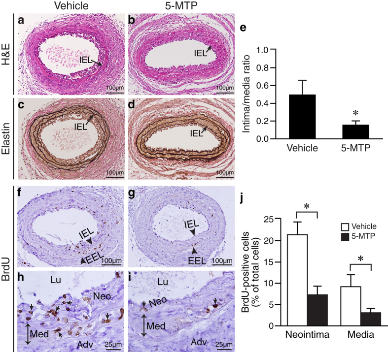
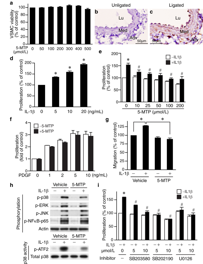
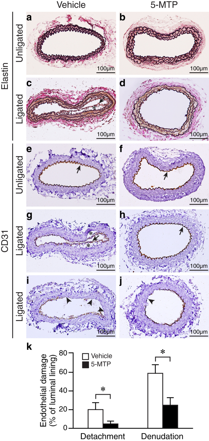
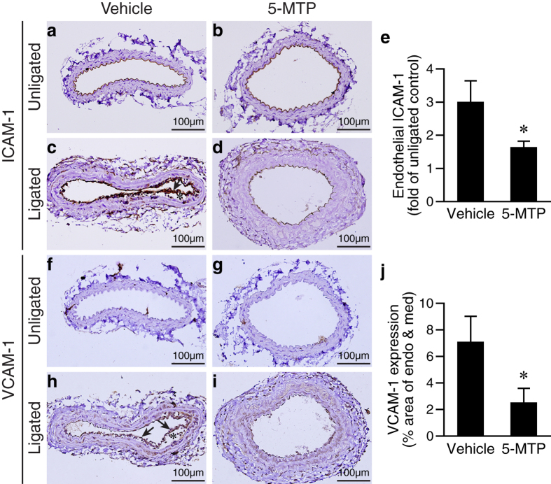
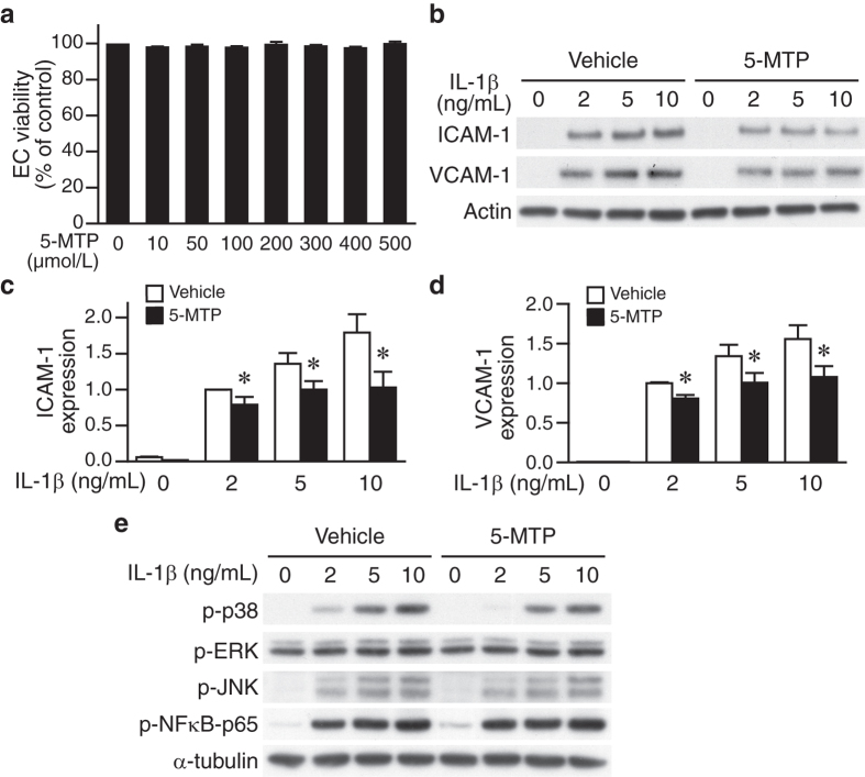
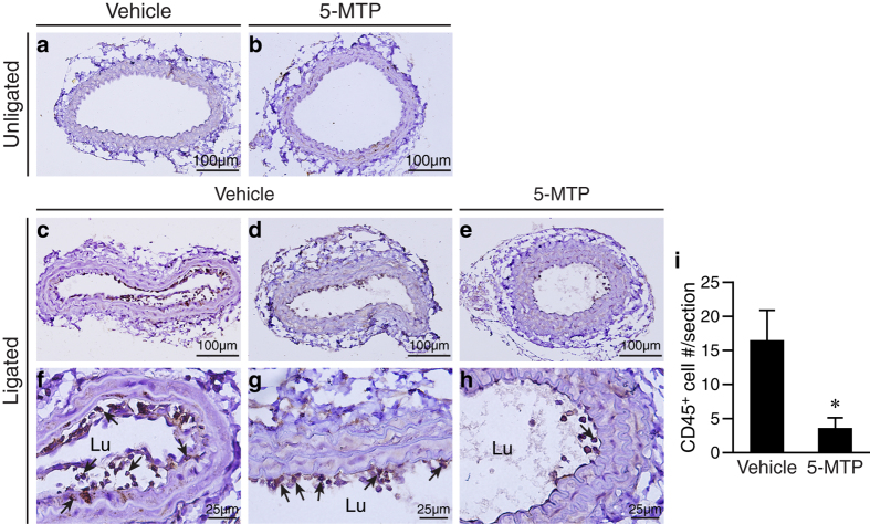
5-Methoxytryptophan (5-MTP), a 5-methoxyindole metabolite of tryptophan metabolism, was recently shown to suppress inflammatory mediator-induced cancer cell proliferation and migration. However, the role of 5-MTP in vascular disease is unknown. In this study, we investigated whether 5-MTP protects against vascular remodeling following arterial injury. Measurements of serum 5-MTP levels in healthy subjects and patients with coronary artery disease (CAD) showed that serum 5-MTP concentrations were inversely correlated with CAD. To test the role of 5-MTP in occlusive vascular disease, we subjected mice to a carotid artery ligation model of neointima formation and treated mice with vehicle or 5-MTP. Compared with vehicle-treated mice, 5-MTP significantly reduced intimal thickening by 40% 4 weeks after ligation. BrdU incorporation assays revealed that 5-MTP significantly reduced VSMC proliferation both in vivo and in vitro. Furthermore, 5-MTP reduced endothelial loss and detachment, ICAM-1 and VCAM-1 expressions, and inflammatory cell infiltration in the ligated arterial wall, suggesting attenuation of endothelial dysfunction. Signaling pathway analysis indicated that 5-MTP mediated its effects predominantly via suppressing p38 MAPK signaling in endothelial and VSMCs. Our data demonstrate a novel vascular protective function of 5-MTP against arterial injury-induced intimal hyperplasia. 5-MTP might be a therapeutic target for preventing and/or treating vascular remodeling.


Figures

Figure 1
Figure 1. Serum 5-MTP concentrations and effect of 5-MTP on mouse blood pressure.
(a) Twenty-three CAD patients and 20 control subjects without CAD and known systemic disease were included in the study. Blood was drawn from patients prior to angiography and serum 5-MTP levels measured by competitive ELISA assays and expressed as μmol/L. *P < 0.0001 vs. control. (b) Approximately 12 weeks old C57BL/6 wild-type mice were injected with 100 mg/kg of 5-MTP by intraperitoneal injection. Blood was collected before (n = 8), after 24 h (n = 4), and 48 h (n = 4) of 5-MTP injections. Serum levels of 5-MTP were measured by competitive ELISA assays. Values are mean ± SE. *P < 0.05 vs. control before exogenous 5-MTP administrations. (c) Systolic blood pressure (SBP) of mice at baseline and at several time points after initial vehicle (n = 6) or 5-MTP (n = 6) injection (100 mg/kg, 3 times a week).
Figure 2
Figure 2. 5-MTP reduces intimal thickening in response to arterial injury.
Mice were subjected to carotid artery ligation, followed by IP injection of vehicle or 5-MTP. Unligated (ad) and ligated (eh) carotid arteries were harvested 4 weeks later. Vessel sections were stained with H&E (a,b,e,f) or Verhoeff’s staining for elastin (black) (c,d,g,h). Representative sections are shown. IEL, internal elastic lamina; EEL, external elastic lamina; neo, neointima. (i) Quantitative morphometric analysis of intimal and medial area in vehicle-treated (n = 9) and 5-MTP-treated mice (n = 12). *P < 0.05 vs. vehicle. (j) Compared with vehicle-treated mice (n = 9), 5-MTP reduced intima/media area ratio (n = 12, *P < 0.05).
Figure 3
Figure 3. 5-MTP inhibits intimal hyperplasia and VSMC proliferation in the injured arteries.
Mice were subjected to carotid artery ligation, followed by IP injection of vehicle (PBS, n = 6) or 5-MTP (n = 9). Carotid arteries were harvested one week after ligation. Mice were injected twice with BrdU at 16–18 h and 1–2 h before harvest. (a,b) Vessel sections were stained with H&E or (c,d) Verhoeff’s staining for elastin (black). Representative sections are shown. IEL, internal elastic lamina. (e) Quantitative morphometric analysis of intimal area/media area ratio in 1-week ligated carotid arteries (*P < 0.05). (fi) Vessel sections were stained with a monoclonal BrdU antibody to detect proliferating cells and counterstained with hematoxylin. Small arrows indicate BrdU-positive brown nuclei. IEL, internal elastic lamina; EEL, external elastic lamina; Neo, neointima; Med, medial layer; Adv, adventitia; Lu, lumen. (j) Immunostained positive cells were quantified as the number of positive cells divided by total number of nuclei in the neointima or media, and expressed as % of total cells. *P < 0.05 vs. vehicle group.
Figure 4
Figure 4. 5-MTP suppresses VSMC proliferation via p38 MAPK and ERK pathway.
(a) MTT assays were performed to assess the effects of 5-MTP on VSMC viability. Immunohistochemistry of (b) unligated and (c) 4 d-ligated vessel sections were performed to detect IL-1β expression (brown). (d) Serum-starved VSMCs were treated with different doses of IL-1β for 24 h, proliferation then assessed by BrdU incorporations and normalized to control without IL-1β stimulation. *P < 0.05 vs. control. (e) Serum-starved VSMCs (in the absence or presence of different doses of 5-MTP) were treated with IL-1β for 24 h and proliferation assessed and normalized to control without IL-1β stimulation. *P < 0.05 vs. control; #P < 0.05 vs. IL-1β-treated but without 5-MTP. (f) Serum-starved VSMCs were stimulated with different concentrations of PDGF-BB in the presence or absence of 5-MTP (100 μmol/L) for 24 h and proliferation assessed. (g) Serum-starved VSMCs (in the absence or presence of 5-MTP) were treated with IL-1β for 24 h, and migration assays performed. VSMCs migrating through the filters were quantified after 4 h. *P < 0.05. (h) Upper panel, VSMCs were serum starved in the absence or presence of 5-MTP, stimulated with or without IL-1β for 15 min, and total proteins isolated for Western blotting to detect phosphorylations of p38 MAPK, ERK1/2, JNK, and NFκB-p65 (Ser536). To verify equal loading, the blots were probed with a pan-actin antibody. A representative of 3 independent experiments is shown. Lower panel, p38 MAPK activity of VSMCs was measured using a p38 MAP kinase assay kit. Phosphorylation of ATF2 indicates p38 MAPK activity. Aliquots of cell lysates were subjected to Western blotting with p38 antibody to verify equivalent amount of protein for activity assessment. A representative of 3 independent experiments is shown. (i) VSMCs were pretreated with p38 MAPK inhibitor SB203580 or SB202190, or MEK inhibitor U0126 30 min prior to IL-1β stimulation. Proliferation was then assessed 24 h later. *P < 0.05 vs. control without IL-1β; #P < 0.05 vs. IL-1β-treated but without inhibitor. Values are mean ± SE of at least three experiments.
Figure 5
Figure 5. 5-MTP protects against endothelial damage of mouse carotid arteries after ligation.
Mice were subjected to carotid artery ligation surgery, followed by IP injection of vehicle (n = 10) or 5-MTP (n = 9). The carotid arteries were harvested 4 d later for histological analysis. (ad) Vessel sections were stained with Verhoeff’s stain for elastin. Unligated vehicle-treated (a) and 5-MTP-treated (b) arteries. (c) Detachment of endothelium (arrow) in the vehicle-treated ligated arteries. *Enlarged subendothelial space. (d) 5-MTP treatment preserves vessel morphology in the ligated arteries. (ej) Vessel sections were stained with endothelial cell marker CD31 antibody to identify endothelium. Arrow indicates endothelium. (g) Detachment of endothelium in the vehicle-treated ligated arteries. *Enlarged subendothelial space. (h) Intact endothelium in the 5-MTP-treated ligated arteries. (i) Loss of endothelium (arrowheads) in the vehicle-treated ligated arteries. (j) Small denuded area (arrowhead) in the 5-MTP-treated ligated arteries. (k) Quantitative morphometric analysis was performed to determine percentage of endothelial detachment and denudation from vehicle- and 5-MTP-treated group (*P < 0.05, n = 5 each group).
Figure 6
Figure 6. 5-MTP suppresses injury-induced adhesion molecule expressions in endothelium.
Vessel sections of carotid arteries harvested 4 d after ligation were stained for adhesion molecule expressions. ICAM-1 expression (brown) in the endothelium of unligated (a,b) and ligated (c,d) arteries. (e) ICAM-1 expression was quantified and expressed as fold of unligated control (*P < 0.05, n = 5 each group). (f,g) VCAM-1 is not detectable in the unligated arteries. (h) Enhanced expression of VCAM-1 in the vessel wall of vehicle-treated ligated arteries. Arrow, endothelium; *subendothelial space. (i) Low level expression of VCAM-1 in the 5-MTP-treated ligated arteries. (j) VCAM-1 expression was measured by colorimetric analysis and expressed as % area of endothelium and media (*P < 0.05, n = 5 each group).
Figure 7
Figure 7. 5-MTP suppresses IL-1β-induced expressions of adhesion molecules and p38 MAPK activation in endothelial cells.
(a) Human umbilical vein endothelial cells (HUVECs) were seeded at a density of 1.6 × 104 cells per well of 96-well plate. After overnight incubation, HUVECs were treated with indicated concentrations of 5-MTP in culture medium for 24 h, and cell viability assessed by MTT assays. (b) HUVECs were pretreated with vehicle or 5-MTP, followed by stimulation with the indicated dose of IL-1β for 24 h. Western blot analyses were performed to detect ICAM-1 and VCAM-1 levels. Blots were subsequently probed with a pan-actin antibody for loading control. A representative of 3 independent experiments is shown. (c) ICAM-1 and (d) VCAM-1 induction is expressed relative to vehicle with 2 ng/mL IL-1β stimulation (because of the very low level expressions without IL-1β stimulation). Values are mean ± SE of 4 experiments. *P < 0.05 vs. vehicle group. (e) HUVECs were pretreated with vehicle or 5-MTP, stimulated with different concentrations of IL-1β for 15 min. Western blotting was performed to detect phosphorylations of p38 MAPK, ERK1/2, JNK, and NFκB-p65 (Ser536). To verify equal loading, the blots were probed with a α-tubulin antibody. A representative of 3 independent experiments is shown.
Figure 8
Figure 8. 5-MTP attenuates vascular injury-induced inflammatory cell adhesion and infiltration.
Mice were subjected to carotid artery ligation, followed by IP injection of vehicle (n = 10) or 5-MTP (n = 9). The unligated (a,b) and ligated (c–h) arteries were harvested 4 d after surgery for histological analysis. Vessel sections were stained with CD45 antibodies for inflammatory cells and representative sections are shown. No CD45-positive cells were detectable in (a) vehicle-treated unligated and (b) 5-MTP-treated unligated arteries. (c) Many CD45-positive (brown color) cells in the vessel wall of vehicle-treated ligated arteries. (d) Many CD45-positive cells on the luminal surface of vehicle-treated ligated arteries. (e) Few CD45-positive cells are detected in 5-MTP-treated ligated vessels. (fh) Higher magnification of (ac), respectively. (f) Arrows indicate CD45-positive cells adhered to the endothelium, infiltrated into subendothelial space, and medial layer. (g) Arrows indicate CD45-positive cells adhered to the luminal surface. (h) Few CD45-positive cells adhered to the luminal surface of ligated arteries from 5-MTP-treated mice. Lu, lumen. (i) Immunostained CD45-positive cells were quantified as the number of positive cells per vessel section. *P < 0.05 vs. vehicle group (n = 5 each group).

Similar articles

Cited by

References

    1. Mozaffarian D. et al.. Heart disease and stroke statistics–2015 update: a report from the American Heart Association. Circulation 131, e29–322, doi: 10.1161/cir.0000000000000152 (2015). - DOI - PubMed
    1. Owens G. K., Kumar M. S. & Wamhoff B. R. Molecular regulation of vascular smooth muscle cell differentiation in development and disease. Physiol. Rev. 84, 767–801 (2004). - PubMed
    1. Doran A. C., Meller N. & McNamara C. A. Role of smooth muscle cells in the initiation and early progression of atherosclerosis. Arterioscler. Thromb. Vasc. Biol. 28, 812–819 (2008). - PMC - PubMed
    1. Ross R. & Glomset J. A. Atherosclerosis and the arterial smooth muscle cell: Proliferation of smooth muscle is a key event in the genesis of the lesions of atherosclerosis. Science 180, 1332–1339 (1973). - PubMed
    1. Donners M. M., Daemen M. J., Cleutjens K. B. & Heeneman S. Inflammation and restenosis: implications for therapy. Ann. Med. 35, 523–531 (2003). - PubMed

Publication types

MeSH terms