

A novel smartphone ophthalmic imaging adapter: User feasibility studies in Hyderabad, India
- PMID: 27146928
- PMCID: PMC4869456
- DOI: 10.4103/0301-4738.181742
A novel smartphone ophthalmic imaging adapter: User feasibility studies in Hyderabad, India
Abstract
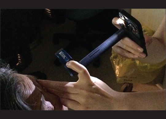
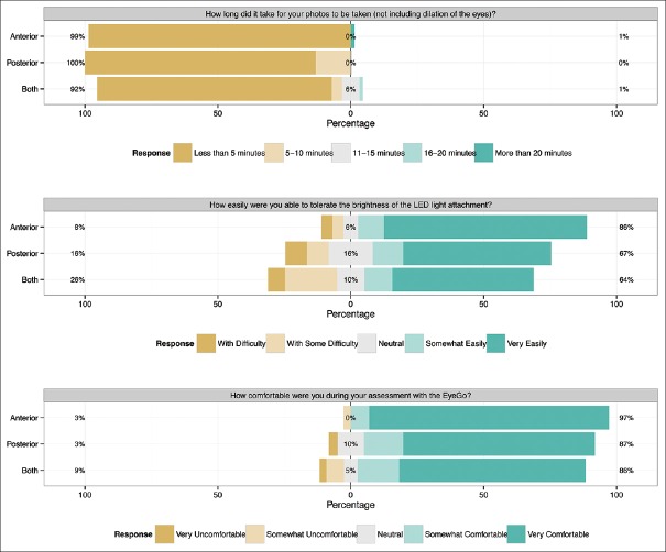
Aim of study: To evaluate the ability of ancillary health staff to use a novel smartphone imaging adapter system (EyeGo, now known as Paxos Scope) to capture images of sufficient quality to exclude emergent eye findings. Secondary aims were to assess user and patient experiences during image acquisition, interuser reproducibility, and subjective image quality.
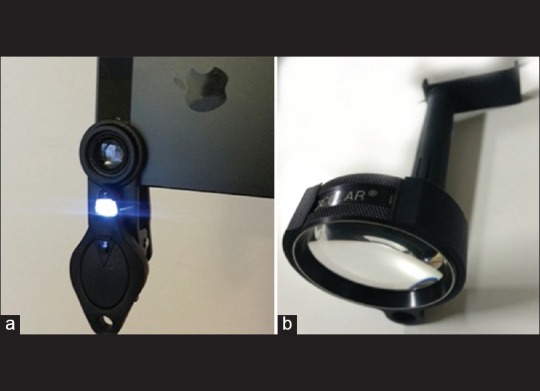
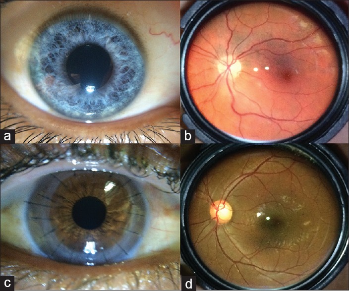
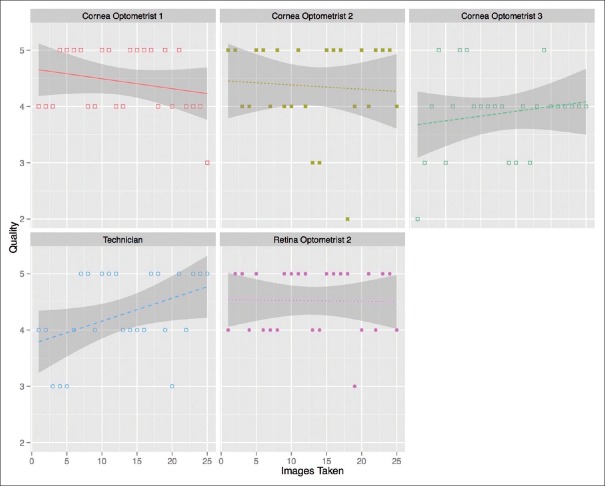
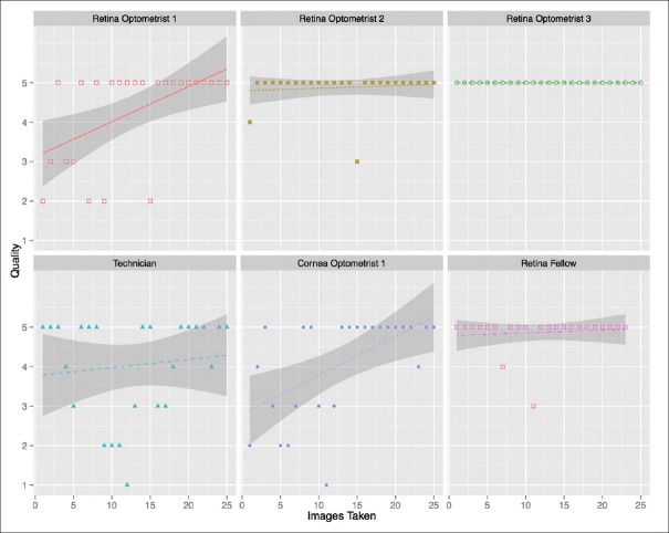
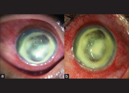
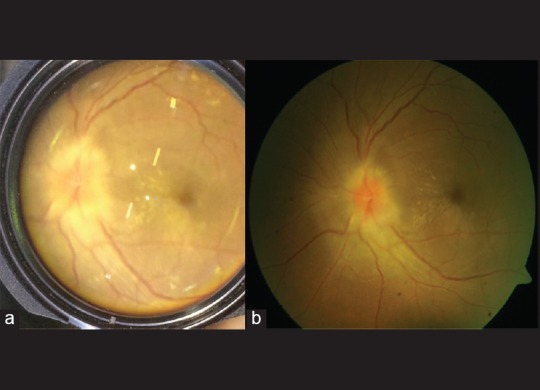
Materials and methods: The system captures images using a macro lens and an indirect ophthalmoscopy lens coupled with an iPhone 5S. We conducted a prospective cohort study of 229 consecutive patients presenting to L. V. Prasad Eye Institute, Hyderabad, India. Primary outcome measure was mean photographic quality (FOTO-ED study 1-5 scale, 5 best). 210 patients and eight users completed surveys assessing comfort and ease of use. For 46 patients, two users imaged the same patient's eyes sequentially. For 182 patients, photos taken with the EyeGo system were compared to images taken by existing clinic cameras: a BX 900 slit-lamp with a Canon EOS 40D Digital Camera and an FF 450 plus Fundus Camera with VISUPAC™ Digital Imaging System. Images were graded post hoc by a reviewer blinded to diagnosis.
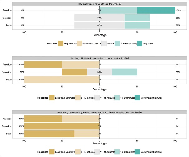
Results: Nine users acquired 719 useable images and 253 videos of 229 patients. Mean image quality was ≥ 4.0/5.0 (able to exclude subtle findings) for all users. 8/8 users and 189/210 patients surveyed were comfortable with the EyeGo device on a 5-point Likert scale. For 21 patients imaged with the anterior adapter by two users, a weighted κ of 0.597 (95% confidence interval: 0.389-0.806) indicated moderate reproducibility. High level of agreement between EyeGo and existing clinic cameras (92.6% anterior, 84.4% posterior) was found.
Conclusion: The novel, ophthalmic imaging system is easily learned by ancillary eye care providers, well tolerated by patients, and captures high-quality images of eye findings.
Figures
References
-
- LVPEI's Mission is to Provide Equitable and Efficient Eye Care to All Sections of Society. Hyderabad, India: L V Prasad Eye Institute; 2015. [Last cited on 2016 Jan 09]. Available from: http://www.lvpei.org/aboutus.php .
-
- Paxos Scope. [Last cited on 2015 Jan 18]. Available from: https://www.digisight.us/digisight/paxos-scope.php .
-
- Myung D, Jais A, He L, Chang RT. Simple, low-cost smartphone adapter for rapid, high quality ocular anterior segment imaging: A photo diary. J Mob Technol Med. 2014;3:2–8.
MeSH terms
Grants and funding
LinkOut - more resources
Full Text Sources
Other Literature Sources
Medical
