Oculodentodigital dysplasia
- PMID: 27146935
- PMCID: PMC4869463
- DOI: 10.4103/0301-4738.180191
Oculodentodigital dysplasia
Abstract
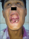
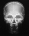
Oculodentodigital dysplasia is a rare, autosomal dominant disorder with high penetrance and variable expressivity, caused by mutations in the connexin 43 or gap junction protein alpha-1 gene. It has been diagnosed in fewer than 300 people worldwide with an incidence of around 1 in 10 million. It affects many parts of the body, particularly eyes (oculo), teeth (dento), and fingers and/or toes (digital). The common clinical features include facial dysmorphism with thin nose, microphthalmia, syndactyly, tooth anomalies such as enamel hypoplasia, anodontia, microdontia, early tooth loss and conductive deafness. Other less common features are abnormalities of the skin and its appendages, such as brittle nails, sparse hair, and neurological abnormalities. To prevent this syndrome from being overlooked, awareness of possible symptoms is necessary. Early recognition can prevent blindness, dental problems and learning disabilities. Described here is the case of a 21-year-old male who presented to the ophthalmology outpatient department with a complaint of bilateral progressive loss of vision since childhood.
Figures


References
-
- Alao MJ, Bonneau D, Holder-Espinasse M, Goizet C, Manouvrier-Hanu S, Mezel A, et al. Oculo-dento-digital dysplasia: Lack of genotype-phenotype correlation for GJA1 mutations and usefulness of neuro-imaging. Eur J Med Genet. 2010;53:19–22. - PubMed
-
- Mubeen K, Vijyalakshmi KR, Ohri N. ODDD – An interesting case. Arch Oral Sci Res. 2011;1:26–9.
-
- Parashari UC, Khanduri S, Bhadury S, Qayyum FA. Radiographic diagnosis of a rare case of oculodentodigital dysplasia. SA J Radiol. 2011;15:134–6.
Publication types
MeSH terms
Supplementary concepts
LinkOut - more resources
Full Text Sources
Other Literature Sources
Medical
Miscellaneous

