. 2016 Aug 1;116(2):456-65.
doi: 10.1152/jn.00857.2015. Epub 2016 May 4.

Knockout of the BK β4-subunit promotes a functional coupling of BK channels and ryanodine receptors that mediate a fAHP-induced increase in excitability

Affiliations

Knockout of the BK β4-subunit promotes a functional coupling of BK channels and ryanodine receptors that mediate a fAHP-induced increase in excitability

Bin Wang et al. J Neurophysiol. .

Abstract

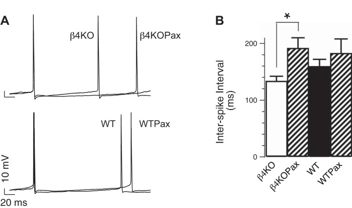
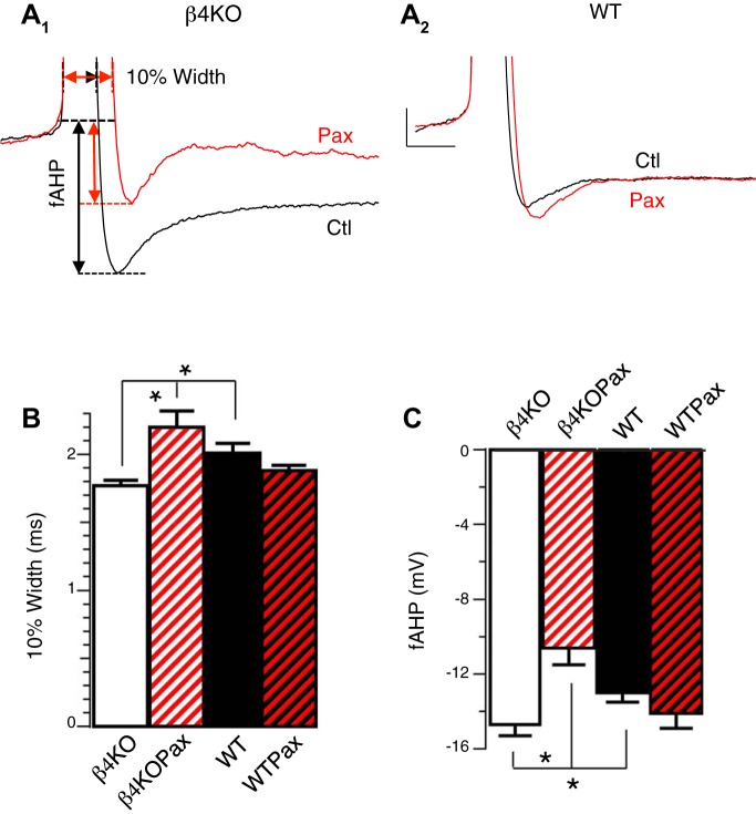
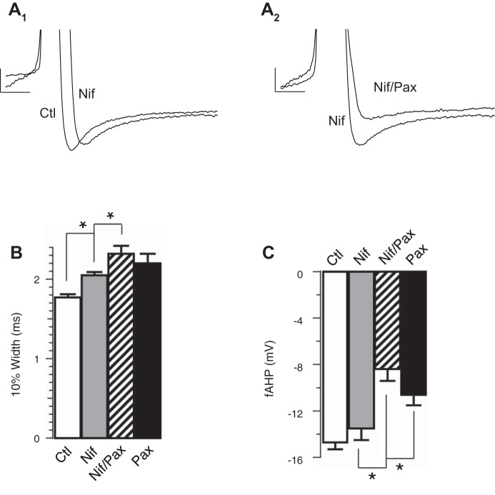
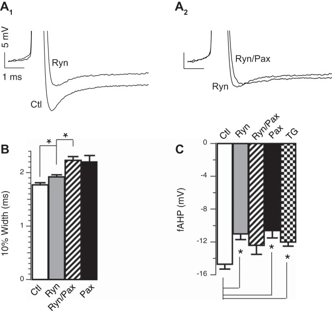
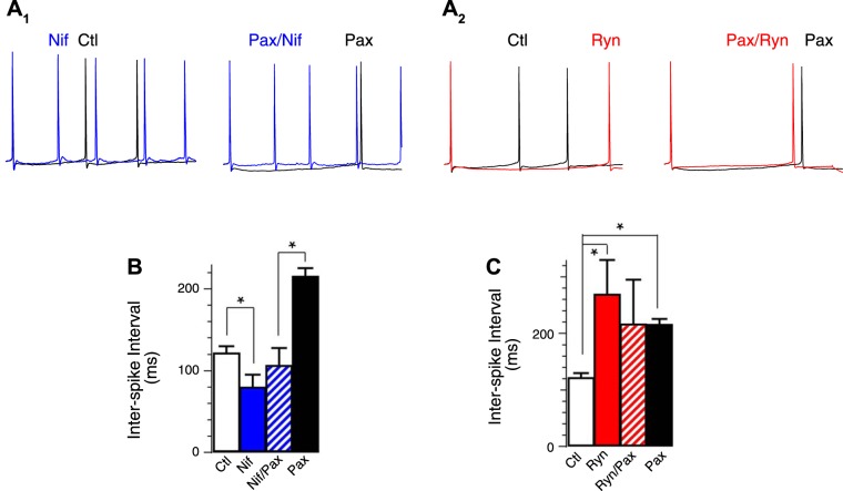
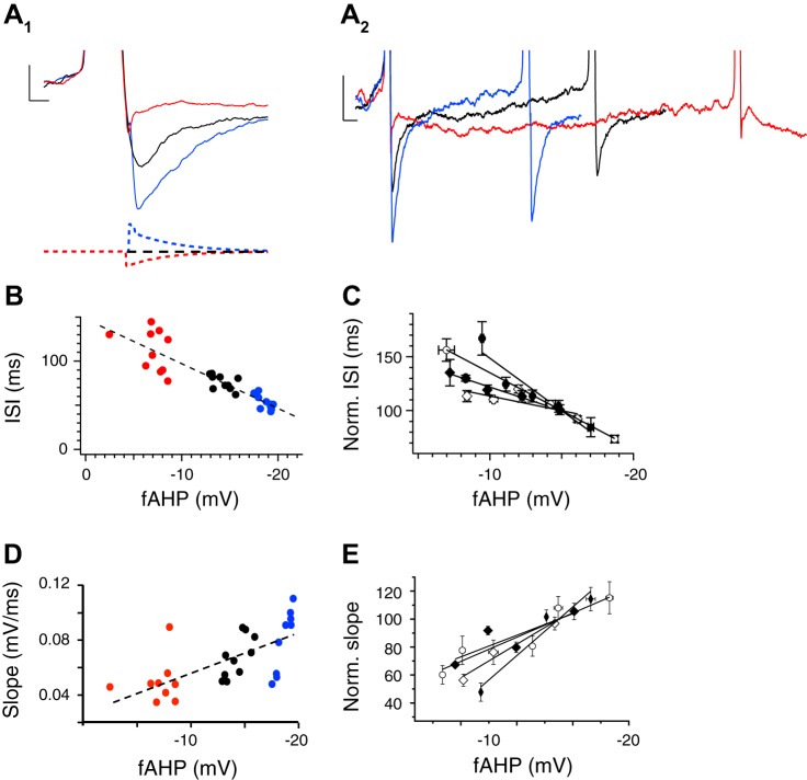
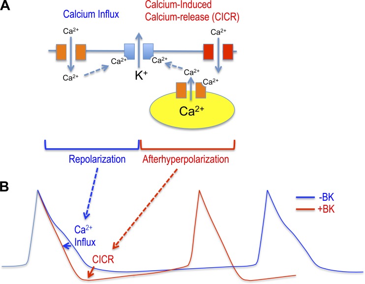
BK channels are large-conductance calcium- and voltage-activated potassium channels with diverse properties. Knockout of the accessory BK β4-subunit in hippocampus dentate gyrus granule neurons causes BK channels to change properties from slow-gated type II channels to fast-gated type I channels that sharpen the action potential, increase the fast afterhyperpolarization (fAHP) amplitude, and increase spike frequency. Here we studied the calcium channels that contribute to fast-gated BK channel activation and increased excitability of β4 knockout neurons. By using pharmacological blockers during current-clamp recording, we find that BK channel activation during the fAHP is dependent on ryanodine receptor activation. In contrast, L-type calcium channel blocker (nifedipine) affects the BK channel-dependent repolarization phase of the action potential but has no effect on the fAHP. Reducing BK channel activation during the repolarization phase with nifedipine, or during the fAHP with ryanodine, indicated that it is the BK-mediated increase of the fAHP that confers proexcitatory effects. The proexcitatory role of the fAHP was corroborated using dynamic current clamp. Increase or decrease of the fAHP amplitude during spiking revealed an inverse relationship between fAHP amplitude and interspike interval. Finally, we show that the seizure-prone ryanodine receptor gain-of-function (R2474S) knockin mice have an unaltered repolarization phase but larger fAHP and increased AP frequency compared with their control littermates. In summary, these results indicate that an important role of the β4-subunit is to reduce ryanodine receptor-BK channel functional coupling during the fAHP component of the action potential, thereby decreasing excitability of dentate gyrus neurons.

Keywords: action potentials; dentate gyrus; fast afterhyperpolarization; large-conductance calcium- and voltage-activated potassium channels.


Figures

Fig. 1.
Fig. 1.
Effect of large-conductance calcium- and voltage-activated potassium (BK) channel blockers on dentate gyrus (DG) action potential (AP) firing. A: representative APs recorded from knockout (KO) β4−/− or wild-type (WT) DG neurons at threshold current injection. Traces represent APs in the absence or presence of 5 μM paxilline (Pax). X and Y scales represent 20 ms and 10 mV, respectively. B: averaged interspike interval (ISI; in ms, β4KO: 133 ± 9, n = 23, β4KOPax: 191 ± 19, n = 8; WT: 159 ± 13, n = 22, WTPax: 182 ± 26, n = 8). β4−/− and β4−/−Pax are significantly different (P = 0.006). Difference between WT and WTPax was not significant (P = 0.4). *P < 0.05.
Fig. 2.
Fig. 2.
β4 Inhibits BK channel function during AP repolarization and fast afterhyperpolarization (fAHP). A: representative APs recorded from β4KO (A1) and WT (A2) DG neurons at threshold current injection in the absence (black) or presence of 5 μM Pax (red). The AP was cut off at the peak to focus on the 10% width and fAHP that were measured as indicated from arrows in A1. B: Pax significantly reduced the 10% AP width in β4KO but not in WT DG. Averaged 10% widths in ms: β4KO 1.77 ± 0.04 (n = 24); β4KOPax 2.20 ± 0.12 (n = 8); WT 2.02 ± 0.06 (n = 29); WTPax 1.88 ± 0.04 (n = 9). WT and β4KO are significantly different (P = 0.004). β4KO and β4KOPax are significantly different (P < 0.001). Difference between WT and WTPax did not reach significance (P = 0.3). C: Pax significantly reduced fAHP amplitude in β4KO but not in WT DG. Averaged fAHP amplitudes in mV: β4KO −14.7 ± 0.6 (n = 24); β4KOPax −10.6 ± 0.9 (n = 8); WT −13.0 ± 0.52 (n = 29); WTPax −14.1 ± 0.8 (n = 9). WT and β4KO are significantly different (P = 0.04). β4KO and β4KOPax are significantly different (P = 0.001). Difference between WT and WTPax was not significant (P = 0.3). *P < 0.05.
Fig. 3.
Fig. 3.
Ca2+ influx through L-type calcium channels affects the repolarization phase. A1: representative threshold APs in the absence or presence of 10 μM nifedipine (Nif) in β4KO neurons. A2: representative threshold APs in the absence or presence of 5 μM Pax in addition to 10 μM Nif. B: Nif increased the 10% width of threshold AP in β4KO neurons. Averaged 10% width in ms: control 1.77 ± 0.04 (n = 24); Pax 2.20 ± 0.12 (n = 8); Nif 2.05 ± 0.04 (n = 11); Nif/Pax 2.32 ± 0.10 (n = 15). Nif and control are significantly different (P ≈ 0.0005). Nif/Pax and Nif are significantly different (P ≈ 0.04). Nif/Pax and Pax are not significantly different (P ≈ 0.46). C: Nif did not reduce the fAHP amplitudes of threshold AP in β4KO neurons. Averaged fAHP amplitudes in mV: control −14.7 ± 0.6 (n = 24); Pax −10.6 ± 0.9 (n = 8); Nif −13.5 ± 1.0 (n = 11); Pax/Nif −8.4 ± 1.0 (n = 15). Nif and control are not significantly different (P ≈ 0.28). Pax/Nif and Pax are not significantly different (P ≈ 0.19). Nif/Pax and Nif are significantly different (P ≈ 0.003). *P < 0.05.
Fig. 4.
Fig. 4.
Ca2+ release through ryanodine receptors (RyR) activates BK during the fAHP. A1: representative threshold APs in the absence or presence of 20 μM ryanodine (Ryn) in β4KO neurons. A2: representative threshold APs in the absence or presence of 5 μM Pax in addition to 20 μM Ryn. B: averaged 10% width in ms: control 1.77 ± 0.04 (n = 24); Pax 2.20 ± 0.12 (n = 8); Ryn 1.92 ± 0.04 (n = 13); Ryn/Pax 2.23 ± 0.07 (n = 9); Ryn and control are significantly different (P ≈ 0.03). Ryn/Pax and Pax are not significantly different (P = 0.82). C: Ryn significantly decreased fAHP amplitudes. Averaged fAHP amplitudes in mV: control −14.7 ± 0.6 (n = 24); Pax −10.6 ± 0.9 (n = 8); Ryn −11.0 ± 0.7 (n = 13); Ryn/Pax −12.4 ± 1.1 (n = 9); thapsigargin (TG) −12.0 ± 0.5 (n = 6). Ryn and control are significantly different (P = 0.0005). Ryn/Pax and Ryn are not significantly different (P = 0.30). Ryn/Pax and Pax are not significantly different (P ≈ 0.24). TG (1 μM) and control are significantly different (P ≈ 0.03). TG and Ryn are not significantly different (P ≈ 0.41). *P < 0.05.
Fig. 5.
Fig. 5.
Nif increased excitability and ryanodine reduced excitability of β4KO neurons. A1 and A2: representative APs in β4KO neurons. The first APs of each trace were aligned. A1: Nif reduced ISI between the 1st and 2nd AP in β4KO neurons. A2: Ryn increased ISI between the 1st and 2nd AP in β4KO neurons. B: averaged ISI in ms, control 121 ± 9 (n = 12); Pax 215 ± 11 (n = 4); Nif 79 ± 16 (n = 8); Nif/Pax 106 ± 22 (n = 5). Nif and control are significantly different (P ≈ 0.02). Nif/Pax is significantly different from Pax (P ≈ 0.005). C: averaged ISI in ms, control 121 ± 9, (n = 12); Pax 215 ± 11 (n = 4); Ryn 268 ± 62 (n = 4); Ryn/Pax 216 ± 80 (n = 5). Pax and control are significantly different (P < 0.0001). Ryn and control are significantly different (P ≈ 0.001). Ryn/Pax is not significantly different from Pax (P ≈ 0.99) or Ryn (P ≈ 0.63). *P < 0.05.
Fig. 6.
Fig. 6.
fAHP amplitude and AP frequency are correlated in β4KO neurons. A1: AP evoked by constant current injection (black trace) is compared with dynamic clamp that increases (blue) and reduces (red) fAHP amplitude. A2: expanded time scale from A1 shows effects of fAHP amplitude on ISIs. B: summary data showing an inverse correlation between fAHP amplitude and ISI for each neuron tested. C: ISI-fAHP relationship for multiple neurons (different symbols for each neuron) normalized to the ISI at the −15 mV fAHP (n = 4). D: summary data showing a correlation between fAHP amplitude and slope (mV/ms) between the fAHP and subsequent spike. The slope is measured from completion of the fAHP of the first spike to threshold of the second spike. E: relative slope-fAHP relationship for multiple neurons (different symbols for each neuron) normalized to the slope at the −15 mV fAHP (n = 4).
Fig. 7.
Fig. 7.
RyR2 gain-of-function mutation R2474S causes an increased fAHP amplitude. A: representative fAHP from R2474S and from control WT sibling dentate gyrus (DG) granule neuron. B and C: fAHP amplitude (control −13.1 ± 0.3; R2474S −16.5 ± 1.0; R2474S + Pax 10.6 ± 1.4, P = 0.004 for control vs. R2474S, P = 0.002 for R2474S vs. R2474S + Pax, n = 8, 9, and 6 respectively, B) and AP 10% width (control 2.0 ± 0.05; R2474S 2.0 ± 0.03; R2474S + Pax 2.2 ± 0.07, P = 0.92 for control vs. R2474S, P = 0.02 for R2474S vs. R2474S + Pax, n = 9, 13 and 6, respectively, C) at threshold current injections. D: ISI at threshold current injections (control 155 ± 43; R2474S 98 ± 10; R2474S + Pax 252 ± 29, P = 0.2 for control vs. R2474S, P = 0.0001 for R2474S vs. R2474S + Pax, n = 8, 8 and 6, respectively). Gray bars are R2474S mice, stippled bars are R2474S + Pax, and black bars are control littermates. *P < 0.05.
Fig. 8.
Fig. 8.
BK proexcitatory effects are mediated by RyR BK channel coupling that increases the fAHP. A: fast-gated BK potassium channels (lacking β4, blue) are activated during the repolarization phase by calcium influx originating from L-type voltage-dependent calcium channels (orange). Different voltage-dependent calcium channels (including T type, red) specifically contribute to calcium-induced calcium release through RyRs (orange) to activate BK channels and increase the fAHP amplitude. B: fAHP, mediated by RyR-BK channel coupling, causes a shorter ISI to confer the BK channel's proexcitatory effect (red trace). Block of BK channels, block of RyRs, or slow-gating conferred by β4 reduces the fAHP amplitude and increases the ISI (blue trace).

Similar articles

Cited by

References

    1. Akita T, Kuba K. Functional triads consisting of ryanodine receptors, Ca(2+) channels, and Ca(2+)-activated K(+) channels in bullfrog sympathetic neurons. Plastic modulation of action potential. J Gen Physiol 116: 697–720, 2000. - PMC - PubMed
    1. Benton MD, Lewis AH, Bant JS, Raman IM. Iberiotoxin-sensitive and -insensitive BK currents in Purkinje neuron somata. J Neurophysiol 109: 2528–2541, 2013. - PMC - PubMed
    1. Berkefeld H, Fakler B. Repolarizing responses of BKCa-Cav complexes are distinctly shaped by their Cav subunits. J Neurosci 28: 8238–8245, 2008. - PMC - PubMed
    1. Berkefeld H, Sailer CA, Bildl W, Rohde V, Thumfart JO, Eble S, Klugbauer N, Reisinger E, Bischofberger J, Oliver D, Knaus HG, Schulte U, Fakler B. BKCa-Cav channel complexes mediate rapid and localized Ca2+-activated K+ signaling. Science 314: 615–620, 2006. - PubMed
    1. Berridge MJ. Neuronal calcium signaling. Neuron 21: 13–26, 1998. - PubMed

Publication types

MeSH terms

LinkOut - more resources