. 2016 May 11;283(1830):20160032.
doi: 10.1098/rspb.2016.0032.

Organic membranes determine the pattern of the columnar prismatic layer of mollusc shells

Affiliations

Organic membranes determine the pattern of the columnar prismatic layer of mollusc shells

Antonio G Checa et al. Proc Biol Sci. .

Abstract

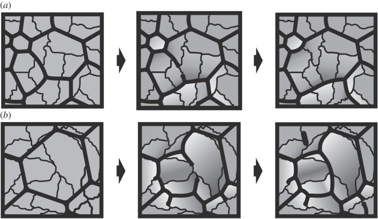
The degree to which biological control is exercised compared to physical control of the organization of biogenic materials is a central theme in biomineralization. We show that the outlines of biogenic calcite domains with organic membranes are always of simple geometries, while without they are much more complex. Moreover, the mineral prisms enclosed within the organic membranes are frequently polycrystalline. In the prismatic layer of the mollusc shell, organic membranes display a dynamics in accordance with the von Neumann-Mullins and Lewis Laws for two-dimensional foam, emulsion and grain growth. Taken together with the facts that we found instances in which the crystals do not obey such laws, and that the same organic membrane pattern can be found even without the mineral infilling, our work indicates that it is the membranes, not the mineral prisms, that control the pattern, and the mineral enclosed within the organic membranes passively adjusts to the dynamics dictated by the latter.

Keywords: biomineralization; mollusc; prismatic layer.


Figures

Figure 1.
Figure 1.
Biogenic (ac) and abiogenic (d) calcite grains with complex morphologies, compared to the polycrystalline prismatic units of pteriomorph bivalves (eh). (a) Tegula funebralis (gastropod). (b) Belemnoid rostrum (Oxfordian, Wittlesey, England). (c) Chama arcana (bivalve). (d) Speleothem (The Pyrenees, locality unknown). (e) Pinctada margaritifera. The inset is a detail of a triple junction between mineral grains. (f) Pinna nobilis. (g) Isognomon legumen. (h) Prismatic pearl of Pinna. The inset shows the organic pellicles which surround the crystalline domains. All surface views. Panels (e,g) have been slightly etched.
Figure 2.
Figure 2.
Electron back scatter diffraction maps of the internal surfaces of Pinctada margaritifera. Secondary electron images and corresponding orientation maps of a wide area (a,b) and two sub-areas indicated in a (cf). The loose membranes are receding membranes. Same specimen as in figure 4a. Different colours indicate different crystal orientations (colour key provided in g). The individual cells contain many crystalline domains and the receding membranes act as boundaries between domains. Note also that the tips of membranes continue into contacts between domains. Upon etching, some crystal boundaries are evident in c,e (arrows).
Figure 3.
Figure 3.
Instances of receding membranes in Pinctada margaritifera. (a) View of the transition of the normal cellular pattern to another pattern composed of wide cells with loose or incomplete membranes. The arrow points towards the shell interior. (b) General view of a similar pattern. Note the abundance of incomplete membranes forming triple junctions with the membrane walls. There are instances of incomplete trifurcate membranes within the cell interiors (arrows). (c,d) Close-up views of two cells with incomplete membranes, some of them about to disappear completely (c). The tips of some of them continue into wavy crystalline boundaries (arrows). (e) Particularly loose pattern. Some of the cells become particularly large (more than 100 µm). Note strongly curved walls (thick arrows). The normal pattern can be seen in the bottom left part of the image. The long thin arrows point towards triple junctions between mineral grains. (f) Semidecalcified specimen showing a receding membrane that finally disappears towards the internal shell surface. Note how the cell wall changes from angled (arrow) to flat with the disappearance of the loose membrane. (g) General view of a completely decalcified specimen showing abundant receding membranes. An instance of a residual nanomembrane that has survived decalcification is indicated (arrow). (h) Details of splitting membranes (arrows) in similarly decalcified specimens. (i) Sketch of the retraction and disappearance of a membrane at a triple junction (thick lines). The receding membrane leaves behind the contact between two crystalline domains (wavy line). With retraction of the membrane, the angle between the other two segments becomes wider, until they form a single flat membrane. Note that Plateau's Law fulfils only for membranes but not for crystalline domains (Cr1, Cr2, Cr3). Panels (ah) are views of the internal shell (growth) surfaces.
Figure 4.
Figure 4.
Instances of advancing membranes in Pinctada margaritifera (ac,h) and pearls of Pinna (dg). (a) General aspect of the external shell surface, with the periostracum removed. Some prismatic units contain two or three crystals (arrows), which are partly fused. (b,c) Instances in which incomplete membranes meet and fuse during shell growth, thus delineating two different prismatic units. In (c), one of the incomplete membranes has rotated and stuck to the cell wall (arrow). (d) Radial fracture through a pearl of Pinna. During growth, some units wedge out (long thin arrow), but the pattern is dominated by the splitting of prismatic units owing to the initiation of organic membranes (thick arrows). (e) Oblique view of the fracture and surface of the same pearl as in (d). The membrane seen on the fracture surface initiates at the position of the white arrow. The tips of the membranes and of their minor branches consistently coincide with boundaries between crystalline domains (black arrows). (f,g) Two views of the growth surface of another pearl. As in (e), the tip of every membrane branch always continues into a nanocrystalline domain boundary. Isolated membranes appear within the cells interior in (f). The arrow in (f) points to the position of a very incipient subsidiary membrane. (h) Locally developed meshwork, possibly delineating individual crystalline domains (arrows point to some crystal boundaries). Panels (ac) are views of the external shell surfaces, and (eh) are views of the growth surfaces.
Figure 5.
Figure 5.
Model for the interaction between membranes and crystals in the CCP layer of bivalves. (a) Sequence for the disappearance of the membrane between two cells thus leading to their complete fusion onto a single one. Recession of the membrane causes the previously separated crystalline domains to come into contact. (b) Sequence for the division of cells owing to production of new membranes. These extend along the boundaries between crystalline domains. In the central cell, two membranes initiate at the opposite ends of a domain until they meet and fuse. Note how angles of triple junctions change with the origination or disappearance of membranes. Light grey indicates contracting crystalline areas and dark grey indicates expanding crystalline areas. See also figure 3i.
Figure 6.
Figure 6.
The Lewis Law formula image 
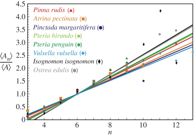
in the CCP layer of bivalves where n is the number of sides of a prism, formula image is the average area of prisms of n sides, formula image is the average area of prisms. The species analysed belong to different orders and superfamilies of the bivalve subclass Pteriomorphia: order Pteroida, superfamily Pinnoidea (Pinna rudis (gradient α = 0.32), Atrina pectinata (α = 0.29)), superfamily Pterioidea (Pinctada margaritifera (α = 0.27), Pteria hirundo (α= 0.33), Pteria penguin (α = 0.34), Vulsella vulsella (α = 0.26), Isognomon isognomon (α = 0.39)), order Ostreoida, superfamily Ostreoidea (Ostrea edulis (α = 0.38)). The relationship is linear for the common prisms with good statistics whose number of sides is close to six.

References

    1. Belcher AM, Wu XH, Christensen RJ, Hansma PK, Stucky GD, Morse DE. 1996. Control of crystal phase switching and orientation by soluble mollusc-shell proteins. Nature 381, 56–58. (10.1038/381056a0) - DOI
    1. Falini G, Albeck S, Weiner S, Addadi L. 1996. Control of aragonite or calcite polymorphism by mollusk shell macromolecules. Science 271, 67–69. (10.1126/science.271.5245.67) - DOI
    1. Mann S. 2001. Biomineralization. Principles and concepts in bioinorganic materials chemistry. Oxford, UK: Oxford University Press.
    1. Weiner S, Dove PM. 2003. An overview of biomineralization processes and the problem of the vital effect. Rev. Mineral. Geochem. 54, 1–29. (10.2113/0540001) - DOI
    1. Cusack M, Freer A. 2008. Biomineralization: elemental and organic influence in carbonate systems. Chem. Rev. 108, 4433–4454. (10.1021/cr078270o) - DOI - PubMed

Publication types

Substances

LinkOut - more resources