. 2016 May 5:6:25255.
doi: 10.1038/srep25255.

Colonization with the enteric protozoa Blastocystis is associated with increased diversity of human gut bacterial microbiota

Collaborators, Affiliations

Colonization with the enteric protozoa Blastocystis is associated with increased diversity of human gut bacterial microbiota

Christophe Audebert et al. Sci Rep. .

Abstract

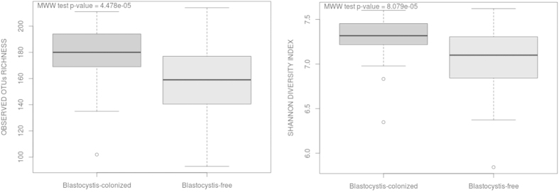
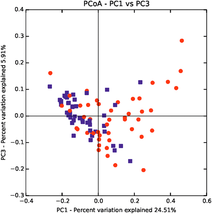
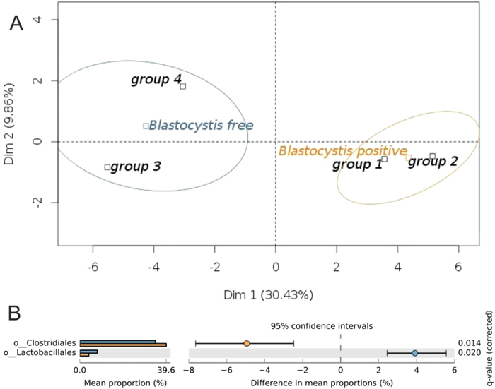
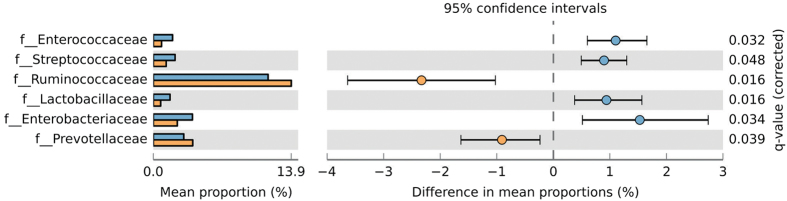
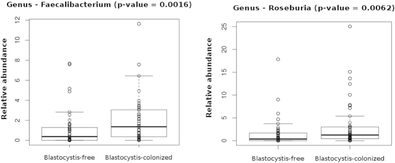
Alterations in the composition of commensal bacterial populations, a phenomenon known as dysbiosis, are linked to multiple gastrointestinal disorders, such as inflammatory bowel disease and irritable bowel syndrome, or to infections by diverse enteric pathogens. Blastocystis is one of the most common single-celled eukaryotes detected in human faecal samples. However, the clinical significance of this widespread colonization remains unclear, and its pathogenic potential is controversial. To address the issue of Blastocystis pathogenicity, we investigated the impact of colonization by this protist on the composition of the human gut microbiota. For that purpose, we conducted a cross-sectional study including 48 Blastocystis-colonized patients and 48 Blastocystis-free subjects and performed an Ion Torrent 16S rDNA gene sequencing to decipher the Blastocystis-associated gut microbiota. Here, we report a higher bacterial diversity in faecal microbiota of Blastocystis colonized patients, a higher abundance of Clostridia as well as a lower abundance of Enterobacteriaceae. Our results contribute to suggesting that Blastocystis colonization is usually associated with a healthy gut microbiota, rather than with gut dysbiosis generally observed in metabolic or infectious inflammatory diseases of the lower gastrointestinal tract.


Figures

Figure 1
Figure 1. Rarefaction curve calculated for Chao1 index demonstrating the higher bacterial diversity found among Blastocystis-colonized patients.
The blue line indicates Blastocystis-colonized patients and the red line indicates Blastocystis-free individuals.
Figure 2
Figure 2. Boxplots of observed OTUs richness and Shannon diversity indexes distinguishing between patients colonized or not by Blastocystis.
Statistical analyses were performed using the Mann-Whitney-Wilcoxon (MWW) test. Plotted are interquartile ranges (IQRs; boxes), medians (dark lines in the boxes), and the lowest and highest values within 1.5 times IQR from the first and third quartiles (whiskers above and below the boxes). Both alpha-diversity metrics were calculated using 273 normalized sequences per sample.
Figure 3
Figure 3. PCoA of the microbial communities in Blastocystis-colonized and Blastocystis-free patient samples.
The blue dots indicate Blastocystis-colonized patients and the red dots indicate Blastocystis-negative individuals.
Figure 4
Figure 4. Analyses of the bacterial microbiota composition at the order-level taxonomic rank.
(A) PCA plot comparing the four patient groups according to their microbiota patterns for the 5 most abundant microbial communities at the order-level taxonomic rank. Groups 3 and 4 define the Blastocystis-free cluster (in blue); groups 1 and 2 define the Blastocystis-positive cluster (in orange). (B) Proportion of sequences assigned to each main group at the order-level taxonomic rank (level 4) for Clostridiales and Lactobacillales illustrated using STAMP, along with means for each group and the significance of the difference in mean proportions using White’s nonparametric t-test with Benjamini-Hochberg FDR multiple test correction. The blue and orange bars represent Blastocystis-free patients and Blastocystis-positive patients, respectively.
Figure 5
Figure 5. Proportion of sequences assigned to each main group at the family taxonomic rank (level 5), along with the means for each group and significance of difference in mean proportions using White’s nonparametric t-test with Benjamini-Hochberg FDR multiple test correction, illustrated using STAMP.
Significant differences (q-value < 0.05) are represented here between the two main groups. Blue and orange bars represent Blastocystis-free patients and Blastocystis-colonized patients, respectively.
Figure 6
Figure 6. Relative abundances of OTUs of Faecalibacterium and Roseburia genera that differ significantly between Blastocystis-colonized and Blastocystis-free patients.
The Mann-Whitney-Wilcoxon test was used to evaluate the two groups.

Similar articles

Cited by

References

    1. Tan K. S. New insights on classification, identification, and clinical relevance of Blastocystis spp. Clin. Microbiol. Rev. 21, 639–665 (2008). - PMC - PubMed
    1. Clark C. G., van der Giezen M., Alfellani M. A. & Stensvold C. R. Recent developments in Blastocystis research. Adv. Parasitol. 82, 1–32 (2013). - PubMed
    1. Bart A. et al. Diagnosis and subtype analysis of Blastocystis sp. in 442 patients in a hospital setting in the Netherlands. BMC Infect. Dis. 13, 389 (2013). - PMC - PubMed
    1. Wawrzyniak I. et al. Blastocystis, an unrecognized parasite: an overview of pathogenesis and diagnosis. Ther. Adv. Infect. Dis. 1, 167–178 (2013). - PMC - PubMed
    1. El Safadi D. et al. Children of Senegal River Basin show the highest prevalence of Blastocystis sp. ever observed worldwide. BMC Infect. Dis. 14, 164 (2014). - PMC - PubMed

Publication types