Review
. 2016 Jul;73(14):2753-64.
doi: 10.1007/s00018-016-2248-4. Epub 2016 May 4.

Starch phosphorylation: insights and perspectives

Affiliations
Review

Starch phosphorylation: insights and perspectives

Sebastian Mahlow et al. Cell Mol Life Sci. 2016 Jul.

Abstract

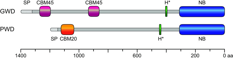
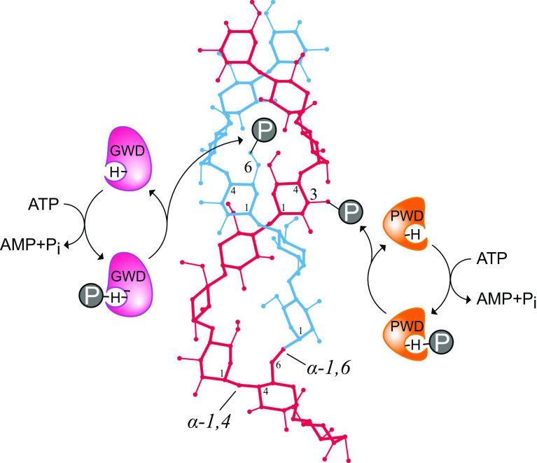
During starch metabolism, the phosphorylation of glucosyl residues of starch, to be more precise of amylopectin, is a repeatedly observed process. This phosphorylation is mediated by dikinases, the glucan, water dikinase (GWD) and the phosphoglucan, water dikinase (PWD). The starch-related dikinases utilize ATP as dual phosphate donor transferring the terminal γ-phosphate group to water and the β-phosphate group selectively to either C6 position or C3 position of a glucosyl residue within amylopectin. By the collaborative action of both enzymes, the initiation of a transition of α-glucans from highly ordered, water-insoluble state to a less order state is realized and thus the initial process of starch degradation. Consequently, mutants lacking either GWD or PWD reveal a starch excess phenotype as well as growth retardation. In this review, we focus on the increased knowledge collected over the last years related to enzymatic properties, the precise definition of the substrates, the physiological implications, and discuss ongoing questions.

Keywords: Glucan, water dikinase; Phosphoglucan, water dikinase; Starch degradation; Starch metabolism; Starch phosphorylation.


Figures

Fig. 1
Fig. 1
Schematic representation of the amino acid sequences of the two plastidial dikinases GWD and PWD from Arabidopsis thaliana including highlighted functional domains. The precursors of both nuclear-encoded dikinase contain an N-terminal signal peptide (SP) for plastid translocation followed by starch-binding domains. In case of GWD, the starch-binding domain is formed by two CBM45s (carbohydrate-binding module; magenta) orientated in tandem domains, which are separated by a 200 amino acid linker. PWD has a single CBM20 (orange) starch-binding domain. The C-terminal part of both sequences shares high homology. It is characterized by a phosphohistidine domain (H*; green) and a nucleotide-binding domain (NB; blue). The unit of the scale at the bottom is amino acids (aa)
Fig. 2
Fig. 2
Schematic illustration of the plastidial dikinases-mediated reactions on α-glucan structures. A left-handed α-1,4-linked glucan chain (red) forms a double helix (upper part) with another glucan chain (blue) connected by a α-1,6-linkage (lower part). Both dikinases act on glucan structures and introduce phosphate esters (P); GWD (magenta) phosphorylates the hydroxyl group at carbon atom 6 while PWD (orange) phosphorylates the C3-hydroxyl group. Before glucan phosphorylation, both enzymes bind and hydrolyze ATP. The γ-phosphate group of ATP is transferred to water and the β-phosphate group to an auto-catalytical histidine residue (H) via a phosphoramidate bond. Subsequently, the β-phosphate is transferred to the glucan substrate
Fig. 3
Fig. 3
Dendrogram of glucan phosphorylating dikinases from land plants and homologs from moss and green algae. Phylogenetic analysis was performed according to [79]. The scale bar indicates amino acid substitutions per site. The amino acid sequences used are according to Joint Genome Institute and as follows: At—Arabidopsis thaliana TAIR10 GWD1 (AT1G10760.1), GWD2 (AT4G24450.1), PWD (AT5G26570.1); Cr—Chlamydomonas reinhardtii v5.5 GWD1 (Cre07.g319300.t1.1), GWD2 (Cre07.g332300.t1.2), PWD1 (Cre17.g719900.t1.2); Os—Oryza sativa v7_JGI GWD1 (LOC_Os06g30310.1), PWD1 (LOC_Os12g20150.1); Ot—Ostreococcus tauri v2.0 GWD1 (118), GWD2 (29353), GWD3 (775), PWD1 (18828), PWD2 (10762); Pp—Physcomitrella patens v3.3 GWD1 (Pp3c8_6520V3.1), GWD2 (Pp3c3_11200V3.1), PWD1 (Pp3c18_14870V3.1), PWD2 (Pp3c14_19150V3.1), PWD3 (Pp3c17_18900V3.1); St—Solanum tuberosum v3.4 GWD1 (PGSC0003DMT400019845), PWD1 (PGSC0003DMT400042818); Vc—Volvox carteri v2.1 GWD1 (Vocar.0003s0059.1), GWD2 (Vocar.0009s0098.1), PWD1 (Vocar.0004s0171.1); Zm—Zea mays Ensembl-18 GWD1 (GRMZM2G412611 _T01), PWD1 (GRMZM2G040968_T01)

References

    1. Buleon A, Colonna P, Planchot V, Ball S. Starch granules: structure and biosynthesis. Int J Biol Macromol. 1998;23(2):85–112. doi: 10.1016/S0141-8130(98)00040-3. - DOI - PubMed
    1. Imberty A, Chanzy H, Perez S, Buleon A, Tran V. The double-helical nature of the crystalline part of a-starch. J Mol Biol. 1988;201(2):365–378. doi: 10.1016/0022-2836(88)90144-1. - DOI - PubMed
    1. Imberty A, Perez S. A revisit to the three-dimensional structure of b-type starch. Biopolymers. 1988;27(8):1205–1221. doi: 10.1002/bip.360270803. - DOI
    1. Deschamps P, Colleoni C, Nakamura Y, Suzuki E, Putaux JL, Buleon A, Haebel S, Ritte G, Steup M, Falcon LI, Moreira D, Loffelhardt W, Raj JN, Plancke C, d’Hulst C, Dauvillee D, Ball S. Metabolic symbiosis and the birth of the plant kingdom. Mol Biol Evol. 2008;25(3):536–548. doi: 10.1093/molbev/msm280. - DOI - PubMed
    1. Ball S. Evolution of the starch pathway. In: Tetlow IJ, editor. Starch: Origins, structure and metabolism. London: The Society for Experimental Biology; 2012. pp. 29–54.

Publication types