Review
. 2016 Jun;13(6):324-37.
doi: 10.1038/nrgastro.2016.59. Epub 2016 May 5.

The Hippo pathway in intestinal regeneration and disease

Affiliations
Review

The Hippo pathway in intestinal regeneration and disease

Audrey W Hong et al. Nat Rev Gastroenterol Hepatol. 2016 Jun.

Abstract

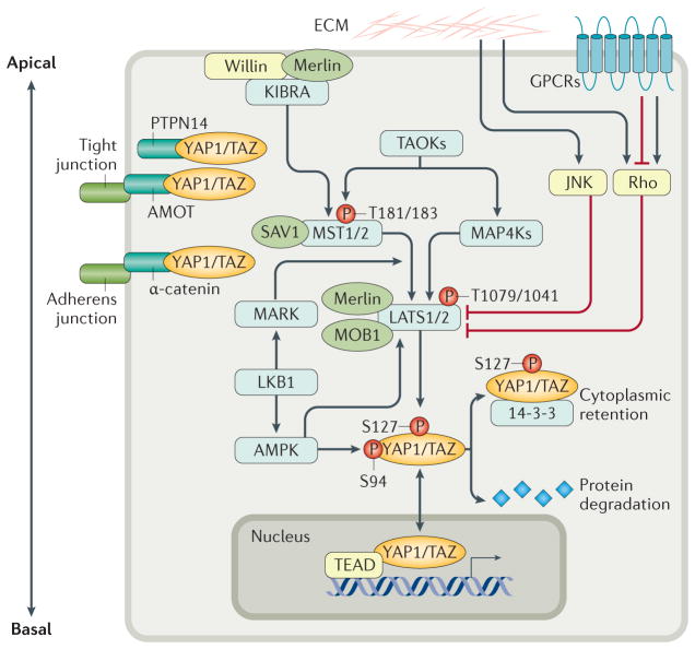
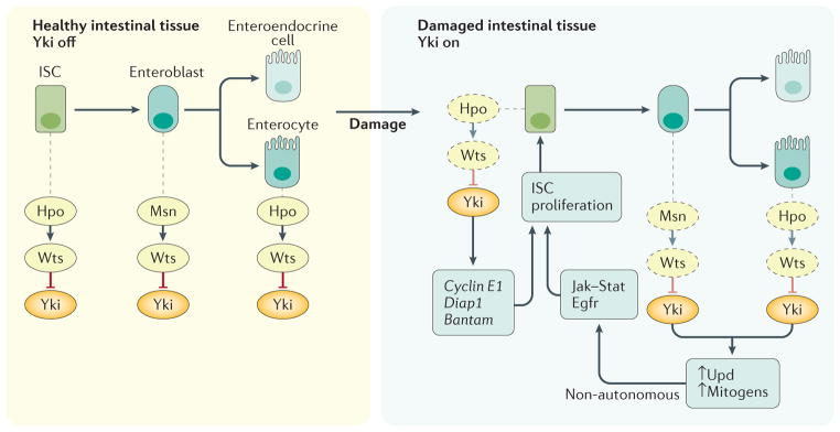
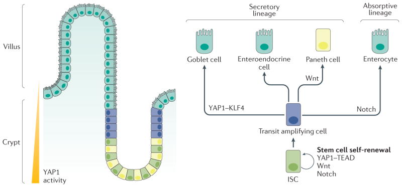
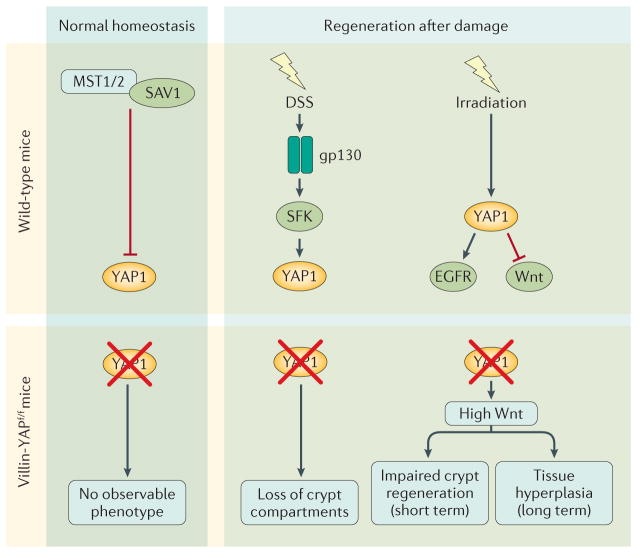
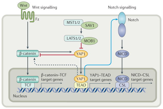
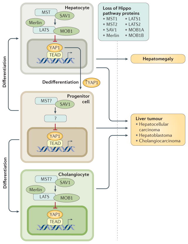
The Hippo pathway is a signalling cascade conserved from Drosophila melanogaster to mammals. The mammalian core kinase components comprise MST1 and MST2, SAV1, LATS1 and LATS2 and MOB1A and MOB1B. The transcriptional co-activators YAP1 and TAZ are the downstream effectors of the Hippo pathway and regulate target gene expression. Hippo signalling has crucial roles in the control of organ size, tissue homeostasis and regeneration, and dysregulation of the Hippo pathway can lead to uncontrolled cell growth and malignant transformation. Mammalian intestine consists of a stem cell compartment as well as differentiated cells, and its ability to regenerate rapidly after injury makes it an excellent model system to study tissue homeostasis, regeneration and tumorigenesis. Several studies have established the important role of the Hippo pathway in these processes. In addition, crosstalk between Hippo and other signalling pathways provides tight, yet versatile, regulation of tissue homeostasis. In this Review, we summarize studies on the role of the Hippo pathway in the intestine on these physiological processes and the underlying mechanisms responsible, and discuss future research directions and potential therapeutic strategies targeting Hippo signalling in intestinal disease.


Conflict of interest statement

Competing interests statement

The authors declare no competing interests.

Figures

Figure 1
Figure 1. The Hippo pathway integrates signals to regulate the activity of YAP1 and TAZ
The core of the Hippo pathway is the kinase cascade of MST1 and MST2 and LATS1 and LATS2. MST proteins, activated by upstream signals, phosphorylate and activate LATS proteins directly, and also activate the scaffold proteins MOB1 and SAV1. MAP4Ks also phosphorylate and activate LATS proteins. Activated LATS proteins phosphorylate YAP1 and TAZ at multiple sites, triggering 14-3-3-mediated cytoplasmic retention and protein degradation. YAP1 and TAZ function as transcriptional co-activators of TEAD proteins to induce expression of genes involved in cell proliferation and apoptosis. Merlin, Willin and KIBRA form a complex to recruit MSTs and LATS to the apical plasma membrane, activating LATS proteins. AMOT and PTPN14 sequesters YAP1 and TAZ at tight junctions, and α-catenin sequesters YAP1 and TAZ at adherens junctions, to prevent their nuclear translocation. LKB1, which has important roles in both cell polarity and cellular stress response, regulates the activity of YAP1 and TAZ through activation of its substrates MARK and AMPK, both of which activate LATS. Activated AMPK also phosphorylates YAP1 and disrupts the YAP1 TEAD complex. The Hippo pathway is further regulated by ECM stiffness and mechanotransduction through Rho GTPases or JNK. GPCRs mediate many extracellular signals to either activate or inhibit LATS via Rho GTPase. AMOT, Angiomotin; AMPK, AMP-activated protein kinase; ECM, extracellular matrix; GPCRs, G protein-coupled receptors; JNK, c-Jun N-terminal kinase; LATS, large tumour suppressor; LKB1, liver kinase B1; MAP4K, mitogen-activated protein kinase kinase kinase kinase; MARK, microtubule affinity-regulating kinase; MOB1, Mob1 homologue; MST, mammalian ste20-like kinase; P, phosphorylation of stated amino acid; PTPN14, protein tyrosine phosphatase non-receptor type 14; SAV1, salvador family WW domain-containing protein 1; TAZ, transcriptional co-activator with PDZ-binding motif; TEAD, TEA domain family member; TAOK, thousand and one amino acid protein kinase; YAP1, Yes-associated protein 1.
Figure 2
Figure 2. The Hippo pathway controls Drosophila midgut homeostasis by restricting Yki activity
In healthy ISCs and enterocytes, the Hpo Wts kinase cascade (homologous to the MST LATS kinase cascade in mammals) suppresses Yki activity. Upon intestinal damage Wts is inactivated, in part by JNK signalling, resulting in Yki activation. Yki activation in enterocytes leads to expression and secretion of Upd protein, a Jak Stat ligand, and Egfr ligands, which promote non-autonomous ISC proliferation in a paracrine manner. Yki activation in ISCs results in expression of cyclin E1, bantam and diap1, promoting autonomous ISC proliferation and survival. In enteroblasts, Msn activates Wts in a Hpo-independent manner, and therefore negatively regulates Yki. Egfr, epidermal growth factor receptor; Hpo, Hippo; ISC, intestinal stem cell; LATS, large tumour suppressor; Msn, misshapen; MST, mammalian ste20-like kinase; Upd, unpaired; Wts, warts; Yki, transcriptional co-activator yorkie.
Figure 3
Figure 3. Mammalian intestine cellular structure and cell fate decision
The mammalian intestinal mucosa is to form villi and crypts. YAP1 activity is highest at the bottom of the crypt, where ISCs and Paneth cells reside. The villus consists of differentiated cells. ISCs in the crypt base differentiate into transit amplifying cells, which further differentiate along a secretory lineage (into goblet cells, enteroendocrine cells and Paneth cells) or an absorptive lineage (into enterocytes). After differentiation, Paneth cells move down to the stem cell niche and reside beside ISCs at the bottom of the crypt. YAP1, Wnt signalling and Notch signalling play important parts in stem cell self-renewal and progenitor cell differentiation. YAP1 interacts with KLF4 to induce progenitor cell differentiation into goblet cells, whereas Wnt signalling is involved in Paneth cell differentiation, and high Notch signalling induces differentiation along the absorptive lineage. ISC, intestinal stem cell; KLF4, krueppel-like factor 4; TEAD, TEA domain family member; YAP1, Yes-associated protein 1.
Figure 4
Figure 4. The Hippo pathway in intestinal homeostasis and injury-induced regeneration
In normal homeostasis, the Hippo pathway is constitutively active to keep YAP1 activity at low levels. Mice lacking YAP1 in intestinal epithelial cells (Villin-YAPf/f mice) have no observable phenotype. During tissue regeneration induced by damage caused by DSS, YAP1 is activated by gp130 signalling to stimulate cell proliferation. Mice with Yap1 deficiency in intestinal epithelial cells have higher mortality and greater loss of crypt compartments than wild-type mice. Whole-body irradiation of mice causes YAP1 activation, which transiently inhibits Wnt and stimulates EGFR signalling to promote regeneration. Loss of YAP1 in intestinal epithelial cells leads to Wnt hyperactivation, impairing crypt regeneration shortly after injury but inducing tissue hyperplasia in the long term. DSS, dextran sulfate sodium; EGFR, epidermal growth factor receptor; gp130, interleukin-6 receptor subunit beta; SAV1, salvador family WW domain-containing protein 1; SFK, Src family kinases; YAP1, Yes-associated protein 1.
Figure 5
Figure 5. Crosstalk between the Hippo pathway and Wnt and Notch signalling in the intestine
When the Hippo pathway is activated, cytoplasmic YAP1 inhibits Wnt signalling by sequestering β-catenin in the cytoplasm. When the Hippo pathway is inactive, nuclear YAP1 upregulates Wnt signalling through an unknown mechanism. In addition, Wnt activation leads to β-catenin-induced activation and upregulation of YAP1. Activated YAP1 transcriptionally upregulates expression of Notch receptors, leading to increased activation of Notch signalling and translocation of the NICD to the nucleus. Notch is considered to be a mediator of the Hippo pathway. Black arrows indicate direct activation, blue arrows indicate transcriptional regulation, dotted lines indicate processes supported by experimental evidence but without a known mechanism. CSL, (CBF1/RBPjκ/Su(H)/Lag-1) transcription factor; Fz, Frizzled; LATS, large tumour suppressor; MOB1, Mob1 homologue; MST, mammalian ste20-like kinase; NICD, Notch intracellular domain; SAV1, salvador family WW domain-containing protein 1; TCF, T-cell factor; TEAD, TEA domain family member; YAP1, Yes-associated protein 1.
Figure 6
Figure 6. Hippo pathway has roles in organ size control and carcinogenesis in different types of liver cells
Disruption of Hippo signalling by deleting core kinase components (MST1, MST2, SAV1, MOB1A, MOB1B, LATS1, LATS2 and Merlin) leads to uncontrolled proliferation of hepatocytes and bile duct epithelial cells (cholangiocytes). Transgenic mice with deficiency in Hippo pathway show spontaneous liver tumours derived from hepatocytes and/or cholangiocytes, as well as hepatomegaly caused by expansion of hepatocytes. Notably, Sav1-knockout mice exhibit expansion of hepatic progenitor cells, which is not observed in mice with deficiency of other Hippo pathway components. On the other hand, induced ectopic expression of YAP1 converts hepatocytes into progenitor or ductal-like cells. These observations indicate a unique role of SAV1 in regulating YAP1 activities in hepatic progenitor roles. LATS, large tumour suppressor; MOB1, Mob1 homologue protein; MST, mammalian ste20-like kinase; SAV1, salvador family WW domain-containing protein 1; YAP1, Yes-associated protein 1.

References

    1. Barry ER, Camargo FD. The Hippo superhighway: signaling crossroads converging on the Hippo/Yap pathway in stem cells and development. Curr Opin Cell Biol. 2013;25:247–253. - PubMed
    1. Piccolo S, Dupont S, Cordenonsi M. The biology of YAP/TAZ: hippo signaling and beyond. Physiol Rev. 2014;94:1287–1312. - PubMed
    1. Harvey KF, Zhang X, Thomas DM. The Hippo pathway and human cancer. Nat Rev Cancer. 2013;13:246–257. - PubMed
    1. Moroishi T, Hansen CG, Guan KL. The emerging roles of YAP and TAZ in cancer. Nat Rev Cancer. 2015;15:73–79. - PMC - PubMed
    1. Yu FX, Meng Z, Plouffe SW, Guan KL. Hippo pathway regulation of gastrointestinal tissues. Annu Rev Physiol. 2015;77:201–227. - PubMed

MeSH terms