. 2016 Jul;263(7):1349-60.
doi: 10.1007/s00415-016-8147-7. Epub 2016 May 4.

Clinical spectrum associated with MOG autoimmunity in adults: significance of sharing rodent MOG epitopes

Affiliations

Clinical spectrum associated with MOG autoimmunity in adults: significance of sharing rodent MOG epitopes

Maria Sepúlveda et al. J Neurol. 2016 Jul.

Abstract

The aim of this study was to report the clinical spectrum associated with antibodies to myelin oligodendrocyte glycoprotein (MOG) in adult patients, and to assess whether phenotypic variants are dependent on recognition of rodent MOG epitopes. We retrospectively analyzed the features, course and outcome of 56 patients whose samples were investigated by brain tissue immunohistochemistry and cell-based assays using human and rodent MOG. The median age at symptom onset was 37 years (range 18-70); 35 patients (63 %) were female. After a median follow-up of 43 months (range 4-554), only 14 patients (25 %) developed a neuromyelitis optica spectrum disorder (NMOSD), 27 patients (47 %) retained the initial diagnosis of isolated optic neuritis, 7 (12 %) of longitudinally extensive transverse myelitis, and 2 (4 %) of acute disseminated encephalomyelitis; 6 patients (11 %) developed atypical demyelinating syndromes (4 had relapsing episodes of short myelitis lesions which in one occurred with optic neuritis; 1 had relapsing brainstem symptoms, and 1 relapsing demyelinating encephalomyelitis). The course was frequently associated with relapses (71 %) and good outcome. Twenty-seven patients (49 %) had antibodies that recognized rodent MOG epitopes, and 9 of them (16 %) showed a myelin staining pattern in rodent tissue. Only the myelin staining pattern was linked to NMOSD (p = 0.005). In conclusion, MOG autoimmunity in adult patients associates with a clinical spectrum wider than the one expected for patients with suspected NMOSD and overall good outcome. Antibodies to rodent MOG epitopes do not associate with any phenotypic variant.

Keywords: Antibodies to myelin oligodendrocyte glycoprotein; Cell-based assays; Immunohistochemistry; Longitudinally extensive myelitis; MRI; Neuromyelitis optica; Optic neuritis.


Conflict of interest statement

Conflicts of interest: Sepúlveda, Armangue, Martinez-Hernandez, Sola-Valls, Sabater, Midaglia, Ariño, Peschl, and Blanco declare that there is no conflict of interest.

Figures

Fig. 1
Fig. 1
Flowchart of the cohort according to the clinical phenotype at onset and at the last follow-up. ADEM acute disseminated encephalomyelitis, LETM longitudinally extensive transverse myelitis, NMOSD neuromyelitis optica spectrum disorder
Fig. 2
Fig. 2
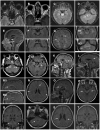
Transverse T2-weighted fluid-attenuated inversion recovery (FLAIR) orbital image shows high signal intensity of both optic nerves (a; arrows), associated with gadolinium enhancement on the T1-weighted transverse image (b; arrows). Transverse T2-FLAIR image shows a central mesencephalic lesion (c; arrow) that extends to the periependymal dorsal pontine region (d; arrow) and shows nodular areas of contrast uptake on the sagittal gadolinium-enhanced T1-weighted image (e; arrows); these brainstem lesions partially resolved on a follow-up MRI performed 6 months latter (f). Transverse T2-FLAIR images show diffuse and confluent white and deep gray matter brain lesions (g; arrows), including the right medial temporal lobe (h1; arrow), the pons, the right middle cerebellar peduncle, and the left dentate nucleus (h2; arrows). Transverse T2-FLAIR image shows lesions affecting the pons, both middle cerebellar peduncles (i; arrows), and the left cerebral peduncle (j; arrow). The same patient shows an extensive corpus callosum lesion on a sagittal T2-FLAIR image (k; arrow), which almost completely resolved 6 months later (l; arrow); a new relapse was associated with a new high signal intensity T2-FLAIR corpus callosum lesion (m1; arrow) that partially enhanced (m2; arrow). Transverse, sagittal, and coronal T2-FLAIR images show multiple patchy areas of increased signal involving the deep white matter and the cortical gray– white junction (n; arrows), the corpus callosum (o; arrow), the midbrain (p; arrows), and the middle cerebellar peduncles (q; arrows); a follow-up at the time of a new relapse showed new pontine periependymal lesions (r; arrow). The same patient shows severe atrophy development and mild diffuse central white matter hyperintensity (s; arrow), but complete resolution of the middle cerebellar peduncle lesions (t; arrows) on transverse and coronal T2-FLAIR images at the last follow-up
Fig. 3
Fig. 3
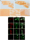
The serum from a patient with human MOG-IgG shows a widespread immunostaining of the white matter (a) in a pattern identical to that obtained with a rabbit polyclonal anti-MOG antibody (b). There is an enhancement of myelin sheaths in the corpus callosum (A1), in pencil fibers of basal ganglia (A2), and in cerebellar white matter (A3). Scale bars 50 μm. HEK293 cells were transfected to express human MOG (c, e green) or mouse MOG (d, f green). Serum from a patient shows antibodies against human (c red) and mouse MOG (d red); serum from a different patient only shows antibodies against human MOG (e red)
Fig. 4
Fig. 4
Serum from a patient shows that the myelin staining pattern in rat brain (a) remains after immunoabsorption with lysates of HEK293 cells transfected with AQP4 as unrelated control (c red) and the reactivity is abrogated (b) when the sample is immunoabsorbed with lysates of HEK293 transfected with mouse MOG (d)

Similar articles

Cited by

References

    1. Mayer MC, Breithaupt C, Reindl M, et al. Distinction and temporal stability of conformational epitopes on myelin oligodendrocyte glycoprotein recognized by patients with different inflammatory central nervous system diseases. J Immunol. 2013;191(7):3594–3604. - PubMed
    1. Reindl M, Di Pauli F, Rostásy K, Berger T. The spectrum of MOG autoantibody-associated demyelinating diseases. Nat Rev Neurol. 2013;9(8):455–461. - PubMed
    1. Pröbstel AK, Dornmair K, Bittner R, et al. Antibodies to MOG are transient in childhood acute disseminated encephalomyelitis. Neurology. 2011;77(6):580–588. - PubMed
    1. Ketelslegers IA, Van Pelt DE, Bryde S, et al. Anti-MOG antibodies plead against MS diagnosis in an acquired demyelinating syndromes cohort. Mult Scler. 2015;21(12):1513–1520. - PubMed
    1. Kitley J, Waters P, Woodhall M, et al. Neuromyelitis optica spectrum disorders with aquaporin-4 and myelin-oligodendrocyte glycoprotein antibodies: a comparative study. JAMA Neurol. 2014;71(3):276–283. - PubMed

MeSH terms

LinkOut - more resources