

Statistical analysis of structural determinants for protein-DNA-binding specificity
- PMID: 27147539
- PMCID: PMC4945413
- DOI: 10.1002/prot.25061
Statistical analysis of structural determinants for protein-DNA-binding specificity
Abstract
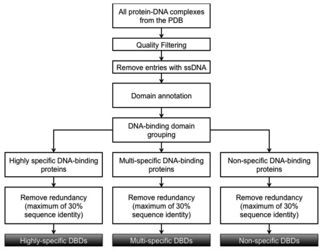
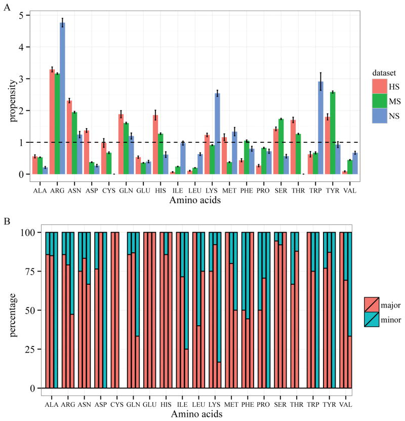
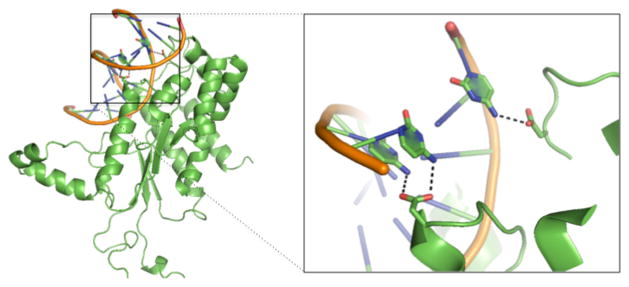
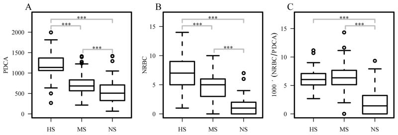
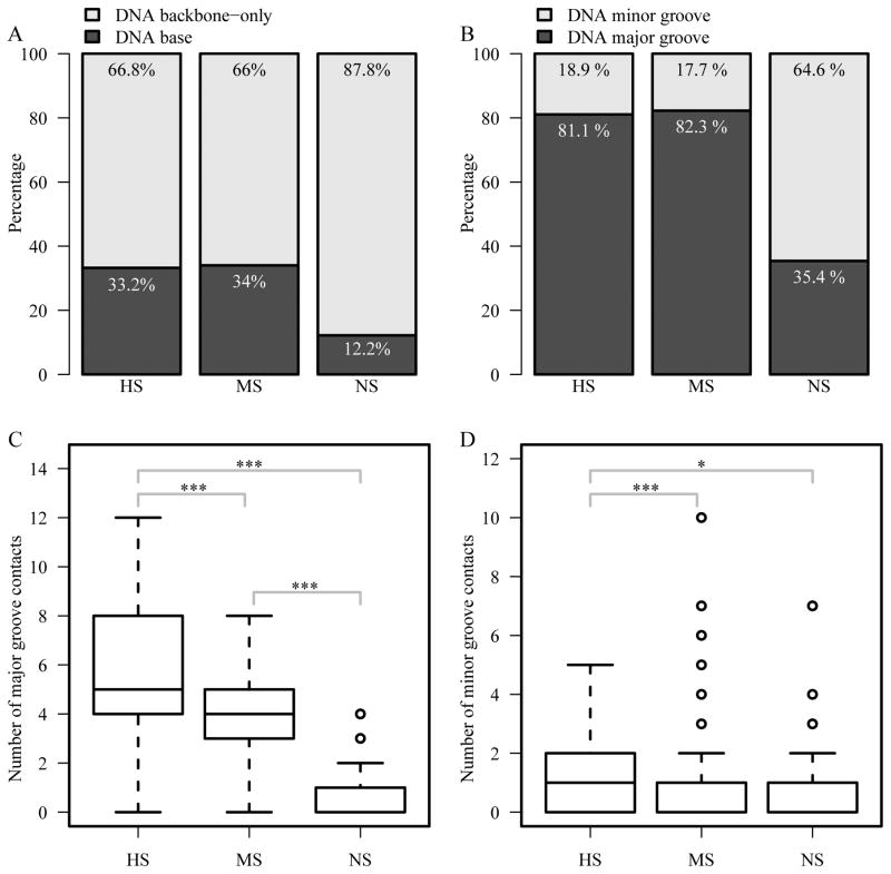
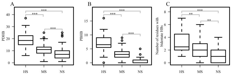
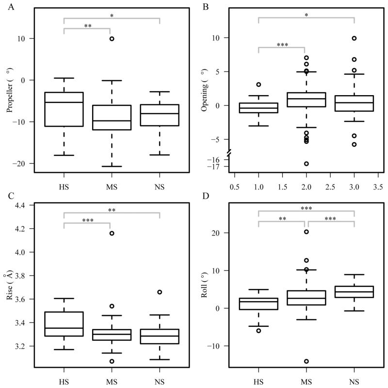
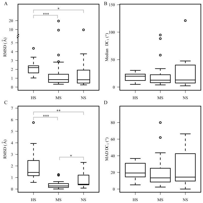
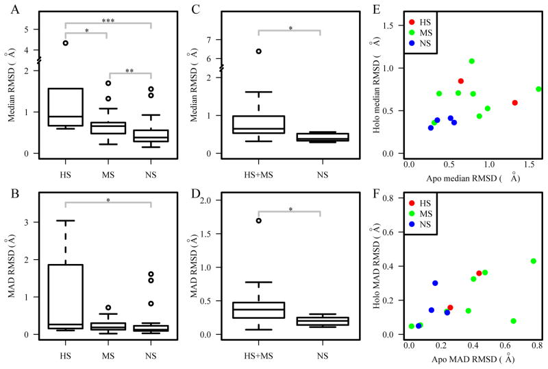
DNA-binding proteins play critical roles in biological processes including gene expression, DNA packaging and DNA repair. They bind to DNA target sequences with different degrees of binding specificity, ranging from highly specific (HS) to nonspecific (NS). Alterations of DNA-binding specificity, due to either genetic variation or somatic mutations, can lead to various diseases. In this study, a comparative analysis of protein-DNA complex structures was carried out to investigate the structural features that contribute to binding specificity. Protein-DNA complexes were grouped into three general classes based on degrees of binding specificity: HS, multispecific (MS), and NS. Our results show a clear trend of structural features among the three classes, including amino acid binding propensities, simple and complex hydrogen bonds, major/minor groove and base contacts, and DNA shape. We found that aspartate is enriched in HS DNA binding proteins and predominately binds to a cytosine through a single hydrogen bond or two consecutive cytosines through bidentate hydrogen bonds. Aromatic residues, histidine and tyrosine, are highly enriched in the HS and MS groups and may contribute to specific binding through different mechanisms. To further investigate the role of protein flexibility in specific protein-DNA recognition, we analyzed the conformational changes between the bound and unbound states of DNA-binding proteins and structural variations. The results indicate that HS and MS DNA-binding domains have larger conformational changes upon DNA-binding and larger degree of flexibility in both bound and unbound states. Proteins 2016; 84:1147-1161. © 2016 Wiley Periodicals, Inc.
Keywords: DNA shape; aromatic residue; aspartate; conformational changes; flexibility; hydrogen bond; structural variations; transcription factor; π-interaction.
© 2016 Wiley Periodicals, Inc.
Figures
Similar articles
-
Conformational changes in DNA-binding proteins: relationships with precomplex features and contributions to specificity and stability.Proteins. 2014 May;82(5):841-57. doi: 10.1002/prot.24462. Epub 2013 Nov 22. Proteins. 2014. PMID: 24265157
-
Major groove recognition by three-stranded beta-sheets: affinity determinants and conserved structural features.J Mol Biol. 2000 Jul 21;300(4):841-56. doi: 10.1006/jmbi.2000.3888. J Mol Biol. 2000. PMID: 10891272
-
Protein-nucleic acid recognition: statistical analysis of atomic interactions and influence of DNA structure.Proteins. 2005 Nov 1;61(2):258-71. doi: 10.1002/prot.20607. Proteins. 2005. PMID: 16121397
-
[The role of weak specific and nonspecific interactions in recognition and conversation by enzymes of long DNA].Mol Biol (Mosk). 2004 Sep-Oct;38(5):756-85. Mol Biol (Mosk). 2004. PMID: 15554181 Review. Russian.
-
What drives proteins into the major or minor grooves of DNA?J Mol Biol. 2007 Jan 5;365(1):1-9. doi: 10.1016/j.jmb.2006.09.059. Epub 2006 Sep 27. J Mol Biol. 2007. PMID: 17055530 Free PMC article. Review.
Cited by
-
Molecular basis for DNA recognition by the maternal pioneer transcription factor FoxH1.Nat Commun. 2022 Nov 26;13(1):7279. doi: 10.1038/s41467-022-34925-y. Nat Commun. 2022. PMID: 36435807 Free PMC article.
-
Improved prediction of DNA and RNA binding proteins with deep learning models.Brief Bioinform. 2024 May 23;25(4):bbae285. doi: 10.1093/bib/bbae285. Brief Bioinform. 2024. PMID: 38856168 Free PMC article.
-
ProDFace: A web-tool for the dissection of protein-DNA interfaces.Front Mol Biosci. 2022 Sep 6;9:978310. doi: 10.3389/fmolb.2022.978310. eCollection 2022. Front Mol Biosci. 2022. PMID: 36148013 Free PMC article.
-
Conformational Change of Transcription Factors from Search to Specific Binding: A lac Repressor Case Study.J Phys Chem B. 2022 Dec 8;126(48):9971-9984. doi: 10.1021/acs.jpcb.2c05006. Epub 2022 Nov 23. J Phys Chem B. 2022. PMID: 36416228 Free PMC article.
-
New insights into protein-DNA binding specificity from hydrogen bond based comparative study.Nucleic Acids Res. 2019 Dec 2;47(21):11103-11113. doi: 10.1093/nar/gkz963. Nucleic Acids Res. 2019. PMID: 31665426 Free PMC article.
References
-
- Schott JJ, Benson DW, Basson CT, Pease W, Silberbach GM, Moak JP, Maron BJ, Seidman CE, Seidman JG. Congenital heart disease caused by mutations in the transcription factor NKX2-5. Science. 1998;281(5373):108–111. - PubMed
-
- Filippova GN, Qi CF, Ulmer JE, Moore JM, Ward MD, Hu YJ, Loukinov DI, Pugacheva EM, Klenova EM, Grundy PE, Feinberg AP, Cleton-Jansen AM, Moerland EW, Cornelisse CJ, Suzuki H, Komiya A, Lindblom A, Dorion-Bonnet F, Neiman PE, Morse HC, 3rd, Collins SJ, Lobanenkov VV. Tumor-associated zinc finger mutations in the CTCF transcription factor selectively alter tts DNA-binding specificity. Cancer research. 2002;62(1):48–52. - PubMed
-
- Luscombe NM, Thornton JM. Protein-DNA interactions: amino acid conservation and the effects of mutations on binding specificity. Journal of molecular biology. 2002;320(5):991–1009. - PubMed
-
- Latchman DS. Transcription-factor mutations and disease. The New England journal of medicine. 1996;334(1):28–33. - PubMed
Publication types
MeSH terms
Substances
Grants and funding
LinkOut - more resources
Full Text Sources
Other Literature Sources
Research Materials
Miscellaneous
