. 2016 Jun 7;7(23):35144-58.
doi: 10.18632/oncotarget.9059.

Prevention effect of rare ginsenosides against stress-hormone induced MTOC amplification

Affiliations

Prevention effect of rare ginsenosides against stress-hormone induced MTOC amplification

Jung-Hyun Cho et al. Oncotarget. .

Abstract

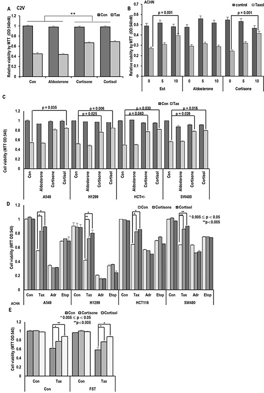
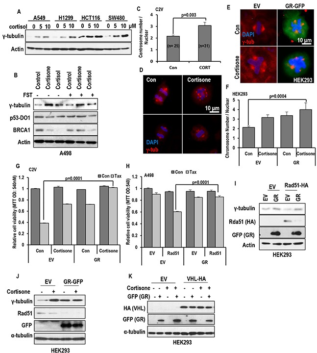
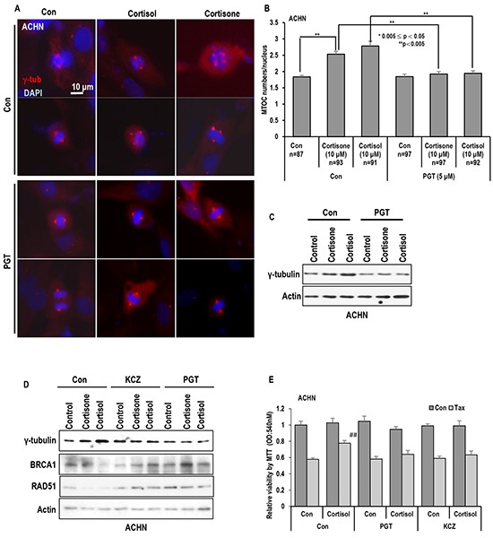
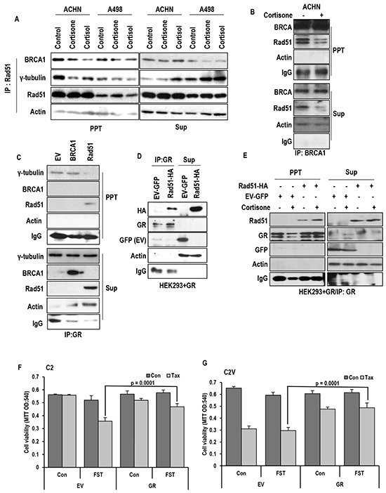
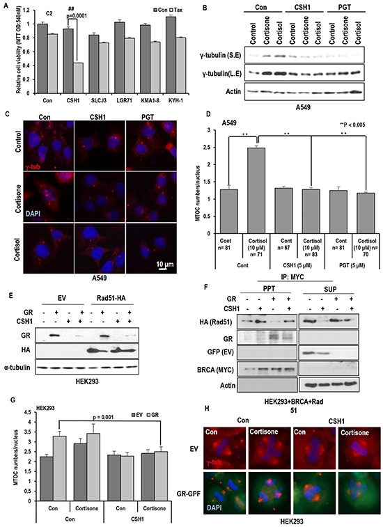
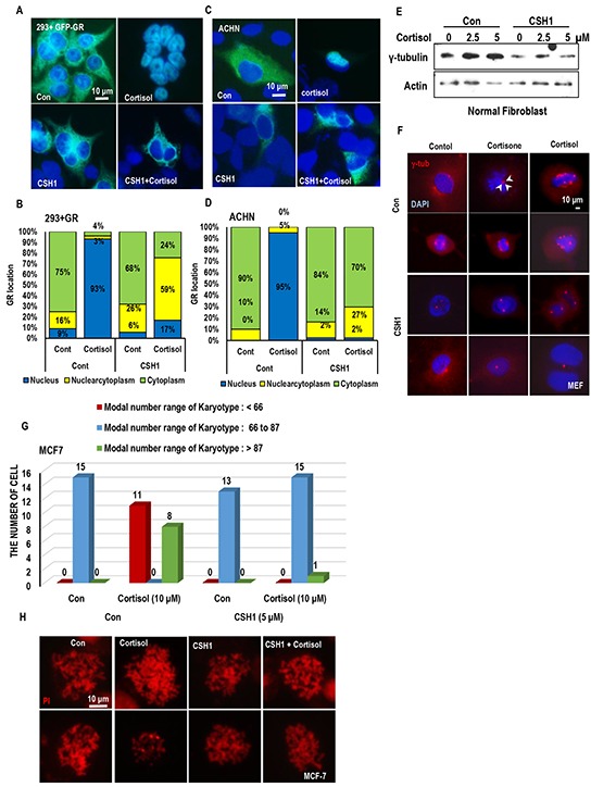
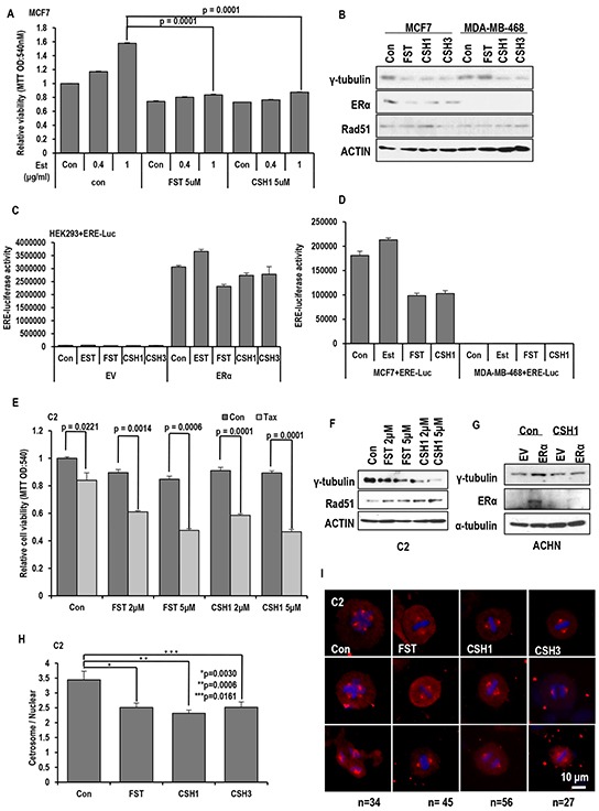
Stress has been suggested as one of important cause of human cancer without molecular biological evidence. Thus, we test the effect of stress-related hormones on cell viability and mitotic fidelity. Similarly to estrogen, stress hormone cortisol and its relative cortisone increase microtubule organizing center (MTOC) number through elevated expression of γ-tubulin and provide the Taxol resistance to human cancer cell lines. However, these effects are achieved by glucocorticoid hormone receptor (GR) but not by estrogen receptor (ER). Since ginsenosides possess steroid-like structure, we hypothesized that it would block the stress or estrogen-induced MTOC amplification and Taxol resistance. Among tested chemicals, rare ginsenoside, CSH1 (Rg6) shows obvious effect on inhibition of MTOC amplification, γ-tubulin induction and Taxol resistance. Comparing to Fulvestant (FST), ER-α specific inhibitor, this chemical can block the cortisol/cortisone-induced MTOC deregulation as well as ER-α signaling. Our results suggest that stress hormone induced tumorigenesis would be achieved by MTOC amplification, and CSH1 would be useful for prevention of stress-hormone or steroid hormone-induced chromosomal instability.

Keywords: MTOC; cancer prevention; stress hormone.


Conflict of interest statement

The authors declare no conflicts of interest.

Figures

Figure 1
Figure 1. Stress hormone induces Taxol resistance
A. Cortisone and Cortisol but not aldosterone block the Taxol-induced cell death. Aldosterone (5 μM), cortisone (5 μM), cortisol (5 μM), and Taxol (3 Mm; Tax) were treated for 72 hr in VHL-positive C2V cells. Cell viability was measured by MTT assay. ** mean different group by ANOVA test (p<0.001). B. Cortisone provides the Taxol resistance, like as Est. ACHN cells were treated with indicating dose of steroid hormones for 72 hr. Cell viability was determined by MTT assay. C. Cortisone and Cortisol but not aldosterone block the Taxol-induced cell death in non-RCC cell lines (Lung cance cell lines; A549, H1299, Colon cancer cell lines; HCT116, SW480). Aldosterone (5 μM), cortisone (5 μM), cortisol (5 μM), and Taxol (3 μM) were treated for 72 hr in VHL-positive C2V cells. Cell viability was measured by MTT assay. D. The specific effect of cortisone on Taxol-induced cell death. Differentially from Taxol (Tax), cortisone did not provide the resistance to DNA damage reagent such as Adriamycin (Adr) and Etoposide (Etop). Cells were incubated with indicated chemicals (Taxol 3 μM; cortisone 5 μM; Adr 2 μg/ml; Etop 10 μM) for 72 hr later. Cell viability was determined by MTT assay. E. Inhibition of ER- α via Faslodex (FST) does not block GH-induced Taxol resistance. ACHN was incubated with FST (3 μM), Taxol (3 μM), cortisone (5 μM) and cortisol (5 μM) for 72 hr. Cell viability was measured by MTT assay.
Figure 2
Figure 2. Stress hormone increases MTOC via GR
A. cortisol obviously induced γ-tubulin in various cell lines. Each cells were treated with indicating dose of cortisol for 72 hr. Western blot was performed for measuring γ-tubulin expression. Actin was used for loading control. B. GHs induce γ-tubulin in regardless of FST. A498 (VHL deficient cell line) were incubated with cortisone (5 μM) and cortisol (5 μM) with/without FST (3 μM) for 24 hr. However, BRCA1 expression was increased by FST, although reduction of BRCA1 expression by GH was not affected. In addition, p53 expression was not altered by FST or GH. Actin was used for loading control. C and D. The number of MTOC is increased by cortisone treatment (CORT; 5 μM) in VHL-intact C2V cells. About 50 cells were counted in each conditions to check average MTOC numbers and representative pictures were shown. Cells were stained with anti-γ-tubulin antibody (Red) to detect MTOC and DAPI (Blue) for DNA. E. GR and cortisone can induce MTOC amplification. After GR transfection or cortisone (5 μM) treatment, HEK293 cells were stained with anti γ-tubulin antibody (Red) and DAPI (Blue). F. Addictive effect of GH and GR on MTOC amplification. About 50 numbers of cells were counted in each condition to estimate MTOC numbers. G. Overexpressed GR can provide the resistance to Taxol in VHL-positive C2V cells. GR was transfected to C2V cells and Taxol (Tax; 3 μM) was treated. 72 hr later, cell viability was measured by MTT assay. H. GR blocks Rad51-mediated Taxol-sensitization. Re-sensitization to Taxol by Rad51 overexpression in VHL-deficient A498 was erased by GR overexpression. A498 cells were transfected with GR and/or Rad51 and incubated with Taxol for 72 hr. Cell viability was determined by MTT assay. I. GR overcomes the Rad51-induced γ-tubulin reduction. In addition, Rad51 is obviously reduced by GR overexpression. GFP-tagged GR and HA-tagged Rad51 were transfected to HEK 293 cells. After 72hr, protein levels were detected with anti-GFP and HA antibodies. J. Obvious reduction of Rad51 by cortisone treatment in GR transfected HEK 293 cells. GFP-tagged GR was transfected to HEK 293 cells and cortisone (5 μM) was treated. After 72hr, western blot was performed. K. GR expression was not altered by VHL. GFP-tagged GR and HA-tagged VHL were transfected to HEK 293 cells and cortisone (5 μM) was treated. After 72hr, western blot was performed. Actin was used as loading control.
Figure 3
Figure 3. Inhibition of GR block the MTOC amplification
A. and B. GR antagonist PGT (Progesterone) blocked the MTOC in ACHN cells. PGT (5 μM) treated cells were blocked MTOC amplication. About 80 cells were counted in each conditions to check average MTOC numbers B and representative pictures were shown A. Cells were stained with anti-γ-tubulin antibody (Red) to detect MTOC and DAPI (Blue) for DNA. C. Treatment of PGT abolished the stress hormone-induced γ-tubulin induction. Western blot analysis was performed with indicated antibodies. Actin was used as loading control. D. Other chemical antagonist of GR, Ketoconazole (KCZ) also blocked the stress hormone effects in ACHN cells. PGT (5 μM) and KCZ (5 μM) were treated. 72 hr. Each protein expression were determined by Western blot. Actin was used as loading control. E. GR antagonist overcome the cortisol induced Taxol-resistance. Cortisol (5 μM), and Taxol (3 μM) were treated for 72 hr in ACHN cells. PGT (5 μM) and KCZ (5 μM) were treated at the same time. Cell viability was measured by MTT assay. ## mean different group from other tested group by ANOVA test (p<0.001).
Figure 4
Figure 4. GR binds to Rad51 and disrupt Rad51-BRCA1 interaction
A. Cortisone (5 μM) and cortisol (5 μM) disrupt binding Rad51-BRCA1 interaction in VHL-positive ACHN cells, but tiny effect on VHL-negative A498 cells. For binding assay, immunoprecipitation (IP) analysis was performed by anti-Rad51 antibody. B. Rad51-BRCA1 binding is reduced by cortisone treatment in VHL-intact ACHN cells. Anti-BRCA1 antibody was used for IP analysis. C. GR only binds to Rad51 but not BRCA1. D. The binding of GR-Rad51 is confirmed by exogenous proteins. GFP-tagged GR and HA-tagged Rad51 were overexpressed in HEK293 cells, and lysates from these cells were used for IP analysis. E. Enhanced binding of Rad51-GR is detected by cortisone (5 μM) treatment. IP analysis was performed by anti-GR antibody. F. Taxol sensitivity induced by FST in VHL-negative C2 cells is blocked through GR overexpression. G. Cortisone induced Taxol resistance is not affected by inhibition of ER-α in VHL-intact C2V cells, which has Taxol sensitivity. After transfection for GR overexpression, Taxol (Tax; 3 μM) and Fulvestant (FST; 3 μM) were treated as indicated. 72 hr later, MTT assay was performed for measuring cell viability.
Figure 5
Figure 5. The effect of Rare ginsenoid on Taxol-induced cell death
A. The effect of various ginsenosides on Taxol-induced cell death. Only CSH1 (Rg6) showed the Taxol sensitization effect in C2. Each ginsenosides (5 μM) and Tax (3 μM) were treated. After72 hr, cell viability was measured by MTT assay. B-D. PGT, CSH1 could abolish the stress hormone-induced γ-tubulin expression in A549 (Lung cancer cell line). PGT (5 μM), CSH1 (5 μM), Cortisol (5 μM) and Cortisone (5 μM) were treated for 72 hr. Western blot was performed for measuring γ-tubulin expression. Actin was used as loading control. Approximately 70 cells were counted for average MTOC numbers and representative pictures are shown. Cells were stained with anti-γ-tubulin antibody (Red) and DAPI (Blue). E. GR expression is suppressed by CSH1. GR induced suppression of Rad51 expression was recovered by CSH1 treatment. F. CSH1 could promote the interaction of BRCA1 and Rad51 and overcome the GR-disrupted binding of them. MYC-tagged BRCA1, HA-tagged Rad51 and GFP-tagged GR were overexpressed in HEK293 cells, and lysates from these cells were used for IP analysis for binding assay. IP analysis was performed by anti-MYC antibody. G and H. CSH1 could block the GR or cortisone induced MTOC amplification. Approximately 50 cells were counted for average MTOC numbers and representative pictures are shown. GFP-tagged GR was transfected to HEK 293 cells. CSH1 (5 μM) and cortisone (5 μM) were treated as indicated. Cells were stained with anti-γ-tubulin antibody (Red) and DAPI (Blue).
Figure 6
Figure 6. CSH1 blocks GR translocation and chromosomal abnormality
A and B. CSH1 blocks GR-translocation in 293 cells. 293 cells were transfected with GFP-GR for 24 hr and incubated with cortisol and CSH1. Representative pictures were provided in A. based on the pictures, cell counting was performed. Nuclear GR (blue bar) in response to cortisol was abolished by CSH1. C and D. The same experiments with A and B were performed with ACHN and obtained the similar results. E. Cortisol induces g-tubulin in normal human fibroblast. Normal fibroblasts (obtained from 81 year-old) were incubated with cortisol for 24 hr. F. MTOC amplification in mouse embryonic fibroblast (MEF) is induced by cortisol and cortisone. MEF, obtained from 13.5 day, were incubated with 5 μM of indicated stress hormone for 72 hr. cells were fixed and stained with γ-tubulin Ab (red). DAPI was used for DNA staining. G and H. CSH1 blocks chromosomal abnormality. MCF-7 (normal chromatin number is 66 to 87) were incubated with stress hormones and CSH1 for 72 hr. Cells were arrested in M-phase by nocodazole and chromosomes were spread out. Number of chrmosomes were counted under fluorescence microscopy.
Figure 7
Figure 7. Similar effect of CSH1 with FST
A. Inhibitory effect of CSH1 on Est-induced cell proliferation. Proliferation of ER-α positive MCF-7 cells was increased by Est-treatment in a dose-dependent manner. Similarly with FST, CSH1 suppresses the Est-induced proliferation in MCF-7. Chemicals were treated as indicated. 72 hr later, MTT assay were performed to check cell viability. B. Suppression of γ-tubulin expression by FST is only in MCF-7 cells, but CSH1 could reduce γ-tubulin in both MCF-7 and MDA-MB-468 cells. C. ER-α mediated transcription activity is reduced by CSH1 and CSH3 in ER-α transfected HEK293 cells. ERE-luciferase vectors and ER-α were co-transfected in HEK 293 cell to estimate ERE-luciferase activity. D. The similar result is reproduced in MCF-7 cells. CSH1 effectively suppressed ERE luciferase activity like FST. ERE-luciferase vectors were transfected to ERα-positive MCF7 cells and ERα-negative MDA-MB-468 cells. ERE- Luciferase assay was performed after the 4 hr treatment of each chemical. E. Taxol-induced cell death is provided by CSH1 similar to FST in VHL-negative C2 cells with dosage-dependent sensitization. F. Western blot analysis shows that expression of γ-tubulin and ER-α are decreased but Rad51 expression is increased by CSH1 treatment. G. The expression of ER-α is suppressed by CSH1. ER-α was transfected to VHL-positive ACHN cells. After the transfection, CSH1 (5 μM) was treated. 72 hr later, western blot was performed. H and I. The MTOC amplification is inhibited by rare ginsenosides compared with FST in VHL-negative C2 cells. About 50 cells were counted for average numbers of MTOC in each indicated conditions and representative pictures are shown. FST (5 μM), CSH1 (5 μM) and CSH3 (5 μM) were treated to VHL-negative C2 cells. After 72 hr, IF staining was performed. Cells were stained with anti-γ-tubulin antibody (Red) and DAPI (Blue).
Figure 8
Figure 8. Summarized diagram
Under stress conditions, GR pathway is activated, response to stress hormone and induces MTOC amplification and chromosomal instability. It would be one of putative tumorigenic mechanism for stress-induced cancer. However, unlike ER- α pathway, GR is not regulated by VHL. Since Rare ginsenoside, CSH1 possesses steroid-like backbone, it blocks GR as well as ER-α pathway-induced MTOC amplification and Taxol sensitivity. So, it would be used as anti-cancer drugs as well as cancer prevention strategy.

Similar articles

Cited by

References

    1. Chida Y, Hamer M, Wardle J, Steptoe A. Do stress-related psychosocial factors contribute to cancer incidence and survival? Nat. Clin. Pract. Oncol. 2008;5:466–475. - PubMed
    1. Sood AK, Bhatty R, Kamat AA, Landen CN, Han L, Thaker PH, Li Y, Gershenson DM, Lutgendorf S, Cole SW. Stress hormone-mediated invasion of ovarian cancer cells. Clin Cancer Res. 2006;15:369–375. - PMC - PubMed
    1. Rudolph KL, Chang S, Lee HW, Blasco M, Gottlieb GJ, Greider C, DePinho RA. Longevity, stress response, and cancer in aging telomerase-deficient mice. Cell. 1999;96:701–712. - PubMed
    1. Johansen C, Olsen JH. Psychological stress, cancer incidence and mortality from non-malignant diseases. Br J Cancer. 1997;75:144–148. - PMC - PubMed
    1. Tsigos C, Chrousos GP. Hypothalamic-pituitary-adrenal axis, neuroendocrine factors and stress. J. Psychosom. Res. 2002;53:865–871. - PubMed

MeSH terms