. 2016 May 4;36(18):4930-9.
doi: 10.1523/JNEUROSCI.2933-15.2016.

Optogenetic Stimulation of Prefrontal Glutamatergic Neurons Enhances Recognition Memory

Affiliations

Optogenetic Stimulation of Prefrontal Glutamatergic Neurons Enhances Recognition Memory

Abigail Benn et al. J Neurosci. .

Abstract

Finding effective cognitive enhancers is a major health challenge; however, modulating glutamatergic neurotransmission has the potential to enhance performance in recognition memory tasks. Previous studies using glutamate receptor antagonists have revealed that the medial prefrontal cortex (mPFC) plays a central role in associative recognition memory. The present study investigates short-term recognition memory using optogenetics to target glutamatergic neurons within the rodent mPFC specifically. Selective stimulation of glutamatergic neurons during the online maintenance of information enhanced associative recognition memory in normal animals. This cognitive enhancing effect was replicated by local infusions of the AMPAkine CX516, but not CX546, which differ in their effects on EPSPs. This suggests that enhancing the amplitude, but not the duration, of excitatory synaptic currents improves memory performance. Increasing glutamate release through infusions of the mGluR7 presynaptic receptor antagonist MMPIP had no effect on performance.

Significance statement: These results provide new mechanistic information that could guide the targeting of future cognitive enhancers. Our work suggests that improved associative-recognition memory can be achieved by enhancing endogenous glutamatergic neuronal activity selectively using an optogenetic approach. We build on these observations to recapitulate this effect using drug treatments that enhance the amplitude of EPSPs; however, drugs that alter the duration of the EPSP or increase glutamate release lack efficacy. This suggests that both neural and temporal specificity are needed to achieve cognitive enhancement.

Keywords: AMPAkine; optogenetics; prefrontal cortex; rat; recognition memory.


Figures

Figure 1.
Figure 1.
Validation of ChR2 expression within the adult rodent mPFC. A, GFP antibody staining showing the expression of the ChR2-YFP fusion protein within the mPFC after unilateral injection of the ChR2 construct (“ChR2” hemisphere). The contralateral hemisphere was injected with PBS and acted as an internal control (“control” hemisphere). White line separates the two hemispheres. The field of view shown (PL/IL) is represented by the striped area on the stereotaxic atlas. B, Antibody staining showing the induction of cFos expression. An increase in the number of cFos+ cells in the ChR2-expressing hemisphere versus the control hemisphere was found after bilateral light stimulation of the mPFC (180.2 vs 75.1 cells/mm2, t test t(2) = −18.43, p = 0.003). C, Activation of ChR2-expressing neurons after light stimulation was confirmed by the colocalization of GFP, NeuN, and cFos (filled arrowheads, 48.1% of GFP-expressing cells). D, ChR2-expressing neurons (filled arrowheads) did not colocalize with GAD67 (outlined arrowheads), indicating the transfection of a non-GABAergic (pyramidal) phenotype (GFP and GAD67 colocalization 0.4%). Scale bars: A, B, 500 μm; C, D, 100 μm.
Figure 2.
Figure 2.
Light stimulation of glutamatergic neurons and OIP discrimination. Light stimulation was delivered to the mPFC immediately after the sample phase during a 5 min delay period. Each animal performed the task twice, once with light stimulation (stim ON) and once without light stimulation (stim OFF), in a fully counterbalanced within-subject design. ChR2-expressing animals showed an increase in discrimination performance (stim × group F(1.0, 11.0) = 11.741, p = 0.006, stim OFF vs stim ON t(5) = −4.55, p = 0.004, n = 7). Light stimulation showed no effect in sham animals (stim OFF vs stim ON, t(5) = 1.29, p = 0.253, n = 6). A significant level of discrimination was shown by all groups except sham animals under light stimulation conditions (#p > 0.05 vs zero). Data shown as mean ± SEM, **p < 0.01 stim OFF vs stim ON.
Figure 3.
Figure 3.
Light stimulation of glutamatergic neurons and NOP discrimination. Light stimulation was delivered to the mPFC immediately after the sample phase during a 5 min delay period. Each animal performed the task twice, once with light stimulation (stim ON) and once without light stimulation (stim OFF), in a fully counterbalanced within-subject design. Discrimination of novel and familiar objects was not affected by mPFC light stimulation in either ChR2 or sham animals. Data are shown as mean ± SEM for sham (n = 6) and ChR2 (n = 7).
Figure 4.
Figure 4.
Light stimulation of glutamatergic neurons and NOL discrimination. Light stimulation was delivered to the mPFC immediately after the sample phase during a 5 min delay period. Each animal performed the task twice, once with light stimulation (stim ON) and once without light stimulation (stim OFF), in a fully counterbalanced within-subject design. Discrimination of novel and familiar locations was not affected by mPFC light stimulation in either ChR2 or sham animals. Data are shown as mean ± SEM for sham (n = 6) and ChR2 (n = 7).
Figure 5.
Figure 5.
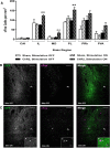
Neuronal activation after light stimulation during the OIP task. A, Light stimulation was delivered to animals that had performed the sample phase of the OIP task. Sham and ChR2 animals were further divided into stim ON or stim OFF groups in a between-subject design. Neuronal activation was increased in ChR2-expressing animals after light stimulation in the PL, IL, and MD regions (IL, p = 0.009; PL, p = 0.009; MD, p = 0.007; stim ON vs stim OFF). B, C, GFP and cFos antibody staining within the PL of ChR2-expressing animals. Light stimulation increased the number of activated ChR2-expressing neurons (C, stim ON 41.5% vs B, stim OFF 18.3%, t(5) = −3.81, p = 0.013), asterisk depicts high-magnification view of GFP and cFos colocalization. Scale bars: C, D, 100 μm; C, high-magnification, 20 μm. Data presented as mean ± SEM for sham stim OFF (n = 3), sham stim ON (n = 3), ChR2 stim OFF (n = 3), and ChR2 stim ON (n = 4). *p < 0.05, **p < 0.01, ChR2 stim OFF versus stim ON.
Figure 6.
Figure 6.
Effect of CX546, CX516, and MMPIP on OIP discrimination. A, Drug infusions were delivered to the mPFC immediately after the sample phase during a 5 min delay period. CX516 improved OIP performance (F(2,14) = 4.95, p = 0.024, 0.3 μg/μl p = 0.008); CX546 and MMPIP showed no effect on discrimination. B, Final injector tip placement of infusion cannula within the mPFC. Injector placements for two animals that were removed due to hemorrhage are not shown. Data are shown as mean ± SEM for CX546 (n = 9), CX516 (n = 8), and MMPIP (n = 10). **p < 0.01 versus vehicle. A significant level of discrimination was shown by all groups except for animals treated with 0.3 μg/μl CX546 (#p > 0.05 vs zero).

References

    1. Arai AC, Xia YF, Rogers G, Lynch G, Kessler M. Benzamide-type AMPA receptor modulators form two subfamilies with distinct modes of action. J Pharmacol Exp Ther. 2002;303:1075–1085. doi: 10.1124/jpet.102.040360. - DOI - PubMed
    1. Aravanis AM, Wang LP, Zhang F, Meltzer LA, Mogri MZ, Schneider MB, Deisseroth K. An optical neural interface: in vivo control of rodent motor cortex with integrated fiberoptic and optogenetic technology. J Neural Eng. 2007;4:S143–156. doi: 10.1088/1741-2560/4/3/S02. - DOI - PubMed
    1. Audet MA, Doucet G, Oleskevich S, Descarries L. Quantified regional and laminar distribution of the noradrenaline innervation in the anterior half of the adult rat cerebral cortex. J Comp Neurol. 1988;274:307–318. doi: 10.1002/cne.902740302. - DOI - PubMed
    1. Barker GR, Warburton EC. NMDA receptor plasticity in the perirhinal and prefrontal cortices is crucial for the acquisition of long-term object-in-place associative memory. J Neurosci. 2008;28:2837–2844. doi: 10.1523/JNEUROSCI.4447-07.2008. - DOI - PMC - PubMed
    1. Barker GR, Warburton EC. When is the hippocampus involved in recognition memory? J Neurosci. 2011;31:10721–10731. doi: 10.1523/JNEUROSCI.6413-10.2011. - DOI - PMC - PubMed

MeSH terms

Substances

LinkOut - more resources