Review
. 2016 Jul 1;311(1):F1-F11.
doi: 10.1152/ajprenal.00166.2016. Epub 2016 May 4.

Pathophysiology of gadolinium-associated systemic fibrosis

Affiliations
Review

Pathophysiology of gadolinium-associated systemic fibrosis

Brent Wagner et al. Am J Physiol Renal Physiol. .

Abstract

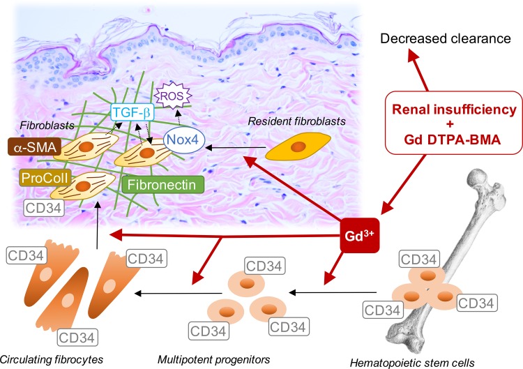
Systemic fibrosis from gadolinium-based magnetic resonance imaging contrast is a scourge for the afflicted. Although gadolinium-associated systemic fibrosis is a rare condition, the threat of litigation has vastly altered clinical practice. Most theories concerning the etiology of the fibrosis are grounded in case reports rather than experiment. This has led to the widely accepted conjecture that the relative affinity of certain contrast agents for the gadolinium ion inversely correlates with the risk of succumbing to the disease. How gadolinium-containing contrast agents trigger widespread and site-specific systemic fibrosis and how chronicity is maintained are largely unknown. This review highlights experimentally-derived information from our laboratory and others that pertain to our understanding of the pathophysiology of gadolinium-associated systemic fibrosis.

Keywords: NADPH oxidase; fibrosis; gadolinium; nephrogenic fibrosing dermopathy; skin diseases.


Figures

Fig. 1.
Fig. 1.
Gadolinium-based contrast: renal clearance and their chemical structures. A: clearance of intravascular contrast. Most contrast agents, whether iodinated or gadolinium based, are eliminated by renal excretion. The concentration of the substance (C) can be determined by a clearance equation. The United States Food and Drug Administration-approved dose of most gadolinium-based contrast is 0.1 mmol/kg. After distribution, with a volume of distribution that is almost entirely the extracellular space, the rapidity of elimination of contrast is determined by renal function. Curves were created based on the reported half-life for magnetic resonance contrast (i.e., the far-right curve) and reducing the rate constant, k, by 20% (modeling the elimination with worsening renal function). B: the various chemical structures of commonly used gadolinium ligands. In general, these can be grouped as being somewhat “linear” or macrocyclic. Note that the number of carboxylic acid side chains determines whether the chelate has a neutral charge (“nonionic”) or not (“ionic”) (3). Vd, volume of distribution.
Fig. 2.
Fig. 2.
Myeloid cells comprise much of the cellularity of gadolinium-based, contrast-induced lesions. A: it has been presupposed that bone marrow-derived circulating cells are the mediators of gadolinium contrast-induced systemic fibrosis. Transgenic rats “tagged” with a human placental alkaline phosphatase antigen served as bone marrow donors to lethally irradiated recipients with 5/6 nephrectomies (as a model of chronic kidney disease). After an engraftment period, one group was treated with gadolinium-based contrast (2.5 mmol/kg intraperitoneally daily, for a total of 20 doses over 4 wk) (101). B: this type of experiment has been conducted several times in our laboratory. Occasionally, animals will demonstrate gross dermatological changes, ranging from hyperpigmentation to punctate ulcerations and eschars. C: histologically, skin changes in rats resembles that found in humans; there is an increase in epidermal thickness, disorganized collagen bundles in the dermis, and an increase in dermal cellularity. D: dermis from the contrast-treated animals demonstrate a marked increase in myeloid cellularity. These cells often express α-smooth muscle actin-positive stress fibers. Immunofluorescence is shown of frozen skin (original magnification ×40). E: immunoblot of fibronectin, a marker of fibrosis, which is increased in the skin from contrast-treated animals. F: the fibrocyte markers procollagen type I (procol) and CD34 are increased in the dermis from contrast-treated animals. This pattern resembles what has been reported in humans afflicted with gadolinium contrast-induced systemic fibrosis. Note that some of the myeloid cells express these markers. DAPI, 4,6-diamidino-2-phenylindole; HPAP, human placental alkaline phosphatase; α-SMA, α-smooth muscle actin.
Fig. 3.
Fig. 3.
Reactive oxygen species mediate gadolinium contrast-induced systemic fibrosis. A: tempol, a superoxide dismutase mimetic, was concomitantly administered (in the drinking water) to rats in a model of contrast-induced fibrosis (101). B: skin from rats treated with magnetic resonance imaging contrast showed dermal fibrosis and an increase in dermal cellularity. This effect was abrogated in the group where tempol was coadministered. Hematoxylin and eosin: top, ×10 objective; bottom, ×40 objective. Calibration bar = 0.05 mm. C: gadolinium contrast-induced skin fibronectin is normalized by coadministration of tempol. D: tempol suppresses the gadolinium-based contrast increases of dermal fibrocyte marker CD34 and α-smooth muscle actin (α-SMA) positive stress fibers. E: magnetic resonance imaging contrast increases skin NADPH oxidase isoform 4 (Nox4) levels. F: animals that received tempol demonstrated less oxidative stress than those subjected to magnetic resonance contrast alone.
Fig. 4.
Fig. 4.
Possible molecular mechanisms underlying gadolinium-based, contrast-induced systemic fibrosis. See text for details.

Similar articles

Cited by

References

    1. Abujudeh HH, Kaewlai R, Kagan A, Chibnik LB, Nazarian RM, High WA, Kay J. Nephrogenic systemic fibrosis after gadopentetate dimeglumine exposure: case series of 36 patients. Radiology 253: 81–89, 2009. - PubMed
    1. Agarwal R, Brunelli SM, Williams K, Mitchell MD, Feldman HI, Umscheid CA. Gadolinium-based contrast agents and nephrogenic systemic fibrosis: a systematic review and meta-analysis. Nephrol Dial Transplant 24: 856–863, 2009. - PubMed
    1. Aime S, Caravan P. Biodistribution of gadolinium-based contrast agents, including gadolinium deposition. J Magn Reson Imaging 30: 1259–1267, 2009. - PMC - PubMed
    1. Anavekar NS, Chong AH, Norris R, Dowling J, Goodman D. Nephrogenic systemic fibrosis in a gadolinium-naive renal transplant recipient. Australas J Dermatol 49: 44–47, 2008. - PubMed
    1. Aoyama T, Paik YH, Watanabe S, Laleu B, Gaggini F, Fioraso-Cartier L, Molango S, Heitz F, Merlot C, Szyndralewiez C, Page P, Brenner DA. Nicotinamide adenine dinucleotide phosphate oxidase in experimental liver fibrosis: GKT137831 as a novel potential therapeutic agent. Hepatology 56: 2316–2327, 2012. - PMC - PubMed

Publication types

MeSH terms

LinkOut - more resources